Keywords
Surfactant, synthetic lung surfactant, surfactant protein B, SP-B peptide mimic, surface activity, oxidation resistance, respiratory distress syndrome, acute respiratory distress syndrome
Surfactant, synthetic lung surfactant, surfactant protein B, SP-B peptide mimic, surface activity, oxidation resistance, respiratory distress syndrome, acute respiratory distress syndrome
Lung surfactant is a lipid-protein mixture that is synthesized by alveolar type II cells and secreted into the alveolus where it reduces surface tension at the air-liquid interface. Mammalian lung surfactant harvested by lavage consists of approximately 80% phospholipids, 10% neutral lipids and 10% protein1. Phospatidylcholine (PC), and particularly dipalmitoylphosphatidylcholine (DPPC), is the major phospholipid constituent of lung surfactant. DPPC enhances the formation of a rigid film at the air-liquid interface that reduces alveolar surface tension to low values during dynamic compression, whereas fluid phospholipids and neutral lipids are important because they significantly improve film spreading2,3. The highly hydrophobic surfactant protein B (SP-B) and, to a lesser extent, surfactant protein C (SP-C), facilitate the absorption of phospholipids into the air-liquid interface and thus play an important role in the reduction of alveolar surface tension. SP-B is pivotal for normal lung function, by hereditary SP-B deficiency being fatal in newborn infants4 and also in SP-B knockout mice5. Human SP-B is a 79 amino-acid, lipid-associating monomer (MW ~8.7 kDa) found in the lung as a covalently linked homodimer. Early theoretical studies based on homology comparisons indicated that the SP-B monomer consists of 4-5 α-helices6–10 with three intramolecular disulfide bridges (i.e., Cys-8 to Cys-77, Cys-11 to Cys-71 and Cys-35 to Cys-46)11, and belongs to the saposin protein superfamily12. The helical bundle for SP-B folds into two leaves, with one leaf having α-helices 1 (N-terminal helix), 5 (C-terminal helix) and 4 and the other composed of α-helices 2 and 313,14.
Intratracheal administration of animal-derived lung surfactants, which contain only polar lipids and native SP-B and SP-C, has greatly improved morbidity and mortality of premature infants with neonatal respiratory distress syndrome (RDS) as a result of surfactant-deficiency due to lung immaturity15. Existing clinically available formulations are extracted from lung lavages or homogenates from pigs (Curosurf®) and cows (Infasurf®, Survanta®), and contain small amounts of SP-B and SP-C (<< 2% of total weight) in a lipid extract with DPPC as its main component. Based on the predicted 3D-saposin motif for SP-B, we have developed minimal SP-B constructs that have desirable structural properties and maintain high activities in animal models of surfactant deficiencies9,10. For example, Super Mini-B (SMB) is a 41-residue, ‘short-cut’ peptide (Figure 1A), based on the primary sequence, secondary structure and tertiary folding of the known sequence of native SP-B (79-residues), that mimics the high surfactant activity of its parent protein10,14,16. SMB incorporates the N-terminal α-helix (~residues 8-25) and C-terminal α-helix (~residues 63-78) of native SP-B as a single linear peptide (Figure 1A), joined together with a customized turn to form a α-helix hairpin. The oxidized SMB in Figure 1A has two vicinal disulfide bonds (i.e., Cys-8 to Cys-77 and Cys-11 to Cys-71) that further covalently link the N- and C-terminal α-helices, and also a hydrophobic N-terminal insertion sequence (i.e., residues 1-7; FPIPLPY). Experimental procedures validated the above structural model for SMB, including conventional 12C-FTIR spectroscopy, mass spectroscopy, I-TASSER homology modeling, and Molecular Dynamics (MD) simulations in lipid mimics and lipid bilayers14,16,17. When formulated with a lipid composition that mimics that of native lung surfactant, SMB has shown excellent surface activity with fresh and stored preparations, which was closely associated with the formation of an α-helix hairpin10,14,17.
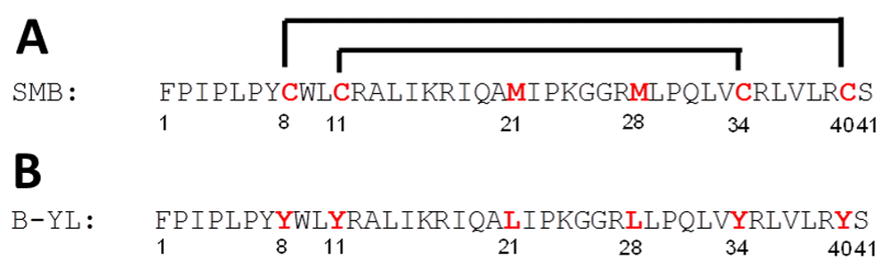
(A) SMB (41 amino-acid residues; 1-letter amino-acid notation), with the N- and C-terminal Phe-1 and Ser-41 as indicated, as well as the sulfur-containing cysteines (Cys-8, Cys-11, Cys-34, Cys-40) and methionines (Met-21, Met-28) in red. The disulfide-linkages are shown between Cys-11 and Cys-34 and between Cys-8 and Cys-40. (B) B-YL (41 residues) shares the same sequence as its parent SMB, except that the cysteines and methionines are replaced by tyrosines (Tyr-8, Tyr-11, Tyr-34, Tyr-40) and leucines (Leu-21, Leu-28) in red, respectively.
Because surfactant therapy is life-saving in preventing and treating RDS in preterm infants, on-going research is studying whether surfactant therapy may be efficaciously extended to pediatric and adult patients with clinical acute lung injury (ALI) or the acute respiratory distress syndrome (ARDS)18. ALI and ARDS may each be caused by direct exposure of lungs to pathogens, oxidative air pollutants, cigarette smoke and other irritants in the alveolar space, and by the presence of endogenous reactive oxygen species (ROS) in damaged lungs due to permeability edema or the inflammatory response. Subsequent oxidative alterations may produce dysfunctional and even inactive lung surfactant in these diseases19,20. SP-B is an important target for ROS-induced oxidative surfactant inactivation21,22. Specifically, oxidation of native SP-B involves alterations in the methionines (Met-29, Met-65) and tryptophan (Trp-9), which correlates well with the loss of in vitro surfactant activity22. Mimics based on the native SP-B sequence may be likewise sensitive to oxidation processes. Kim et al.23 reported that ozone treatment of SP-B(1–25), an SP-B mimic whose sequence overlaps residues 1–25 of SMB (Figure 1A) and native SP-B, variably oxidized amino-acids known to react with ozone. In contrast to the nearly complete homogenous oxidation of the susceptible SP-B(1–25) residues (i.e., Cys-8, Cys-11, Trp-9, and Met-21) in the solvent phase, only a limited subset of residues (Trp-9 and Met-21) oxidized in the hydrophobic interfacial environment provided by the lipid surfactant layer23. In additional studies, Hemming et al.24 showed that exposure of either SP-B1–25 or SMB at the air-water interface to dilute ozone (~2 ppm) produced a rapid loss of surface activity (i.e., increase in surface tension). Because decreases in tryptophan fluorescence occurred concurrently with increasing surface tension for these two SP-B mimics24, it is likely that oxidative disruption of the indole ring of Trp-9 plays a role in the diminished surface activity, possibly due to a fraying of the N-terminal α-helix24,25.
The development of surfactant therapy for ALI/ARDS is especially challenging as it requires the use of exogenous surfactants that successfully resist inhibition from endogenous ROS in injured lungs. Synthetic lung surfactant with SP-B and SP-C peptide mimics offers substantial advantages over current animal-derived surfactants for treating surfactant deficiency in neonatal RDS and surfactant dysfunction in ALI/ARDS. Current research on synthetic lung surfactant has focused on designing peptide mimics of natural surfactant proteins that are highly effective, stable, and easy to manufacture9,10. Here, we conducted structural and functional experiments on ‘B-YL’ (Figure 1B), a 41-residue SMB variant that has its four Cys and two Met residues replaced by Tyr (Tyr-8, Tyr-11, Tyr-34 and Tyr-40) and Leu (Leu-21 and Leu-28), respectively, and tested whether these hydrophobic substitutions produce a surface-active, α-helix hairpin.
HPLC grade chloroform, methanol, trifluoroethanol (TFE), and acetonitrile were purchased from Fisher Scientific (Pittsburgh, PA 15275), trifluoroacetic acid from Sigma Chemical Co (Saint Louis, MO 63103), NMR quality deuterated water was from Aldrich Chemical Co. (St. Louis, MO 63103), and Sephadex LH-20 chromatography gel from Pharmacia (Uppsala, Sweden). Phospholipids were supplied by Avanti Polar Lipids (Alabaster, AL 35007), and Sodium Dodecyl Sulfate (SDS) detergent was from Sigma Chemical Co (Saint Louis, MO 63103).
The oxidized Super Mini-B (SMB) peptide sequence (Figure 1A) was synthesized using a standard Fmoc protocol with a Symphony Multiple Peptide Synthesizer (Protein Technologies, Inc., Tucson, AZ 87514) or a CEM Liberty microwave synthesizer (CEM Corporation, Mathews, NC 28104), cleaved-deprotected and purified using reverse phase HPLC as described earlier14,17. This synthesis protocol included folding of the peptide in a structure-promoting TFE-buffer solvent system to promote oxygen-mediated disulfide linkages between Cys-8 and Cys-40 and between Cys-11 and Cys-3410,14. This covalently stabilized connectivity gave the peptide a helix-hairpin conformation, comparable to the topological organization seen for the N- and C-terminal helical domains of the saposin family of proteins9,10,12. The synthesis of B-YL was identical to that of SMB, except for replacing cysteines with tyrosines (Tyr-8, Tyr-11, Tyr-34, and Tyr-40) and methionines with leucines (Leu-21 and Leu-28), and also omitting the oxidation step. The purified SMB and B-YL peptides were each freeze-dried directly, and the masses were confirmed by MALDI TOF mass spectrometry as described previously17.
Peptide and lipids were formulated as lipid-peptide dispersions to have a total of 2–4% by mole fraction of SMB or B-YL and 35 mg of total lipid [i.e., DPPC: POPC: POPG 5:3:2 mole:mole:mole] per mL of dispersion. The peptide was dissolved in 10 mL of trifluoroethanol and co-solvated with the lipid in chloroform, followed by removal of the solvents with a stream of nitrogen gas and freeze drying of the resulting lipid-peptide film to remove residual solvent. The film was then dispersed with Phosphate Buffered Saline and the sample flask containing the hydrated film was rotated for 1 h at 60°C to produce a solution of multilamellar vesicles (MLVs)14. Lipid controls were similarly prepared but without peptide. These dispersions were then stored at 4°C prior to structural and functional measurements. To determine the molecular mass of peptides formulated with lipids, the peptide was separated from lipid using normal phase chromatography with Sephadex LH-2026.
CD spectra (190–260 nm) of the B-YL peptide in various structure-promoting environments, including surfactant dispersions, were measured with a JASCO 715 spectropolarimeter (Jasco Inc., Easton MD 21601). The instrument was routinely calibrated for wavelength and optical rotation using 10-camphorsulphonic acid27. The sample solutions were scanned using 0.01 cm pathlength cells at a rate of 20 nm per minute, a sample interval of 1 nm, and a temperature of 37°C. Sample concentration was determined by UV absorbance at 280 nm28. Peptide concentration was 100 μM in sample solutions with either TFE:Phosphate buffer (10 mM, pH 7.0) having a volume ratio of 4:6 (v/v), SDS micelles (100 mM) in phosphate buffer (10 mM, pH 7.0), or Single Unilamellar Vesicles (SUVs) of simulated surfactant lipids (DPPC: POPC: POPG; 5:3:2, mole:mole:mole). Surfactant lipid SUVs were prepared at a concentration of 2.6 μM lipids/mL of phosphate buffer solution (10 mM, pH 7.0) by bath sonication for 10 minutes (https://avantilipids.com/tech-support/liposome-preparation/). Sample spectra were baseline corrected by subtracting spectra of protein-free solution from that of the protein-bound solution and expressed as the Mean Residue Ellipticity [θ]MRE as shown in equation (1):
The symbol θ is the measured ellipticity in millidegrees, l is the pathlength in cm, N is the number of residues in the peptide, and c is the concentration of the peptide in mM.
Quantitative estimates of the secondary structural contributions were also made with SELCON 329 using the spectral basis set for membrane proteins, option 4 implemented from the DichroWeb website30,31.
ATR-FTIR spectra were recorded at 37°C using a Bruker Vector 22 FTIR spectrometer (Pike Technologies, Fitchburg, WI 53719) with a deuterium triglyceride sulfate (DTGS) detector. The spectra were averaged over 256 scans at a gain of 4 and a resolution of 2 cm-114. For spectra of B-YL and SMB in TFE solutions, self-films were first prepared by air-drying peptide originally in 100% HFIP onto a 50 × 20 × 2 mm, 45° attenuated total reflectance (ATR) crystal for the Bruker spectrometer. The dried peptide self-films were then overlaid with solutions containing 40% TFE/60% deuterated-10 mM sodium phosphate buffer (pH 7.4), at a peptide concentration of 470 μM. Control solvent samples were similarly prepared for FTIR analysis, but without peptide. Spectra of peptides in solvent were obtained by subtraction of the solvent spectrum from that of peptide solvent. For FTIR spectra of B-YL and SMB in either SDS micelles or surfactant lipids, each lipid-peptide solution was transferred onto a germanium ATR crystal. The aqueous solvent was then removed by flowing nitrogen gas over the sample to produce a thick lipid-peptide (lipid:peptide ratios of 10:1, mole:mole)14. The multilayer film was then hydrated to ≥35% with deuterated water vapor in nitrogen for 1 h before acquiring the spectra32. The spectra for either the B-YL or SMB peptides in the film were obtained by subtracting the spectrum of a peptide-free control sample from that of the peptide-bound sample. The relative amounts of α-helix, β-turn, β-sheet, or random (disordered) structures in lipid-peptide films were estimated using Fourier deconvolution (GRAMS AI 8, version 8.0, Thermo Fisher Scientific, Waltham, MA 02451). The respective areas of component peaks were calculated using curve-fitting software (Igor Pro, version 1.6, Wavemetrics, Lake Oswego, OR 97035)33. FTIR frequency limits were: α-helix (1662-1650 cm-1), β-sheet (1637-1613 cm-1), turn/bend (1682-1662 cm-1), and disordered or random (1650-1637 cm-1)34.
Preliminary structural models for SMB and B-YL were determined by analyzing the respective amino acid sequences with a recent homology modeling program35,36. The homology three-dimensional (3D) structure for oxidized SMB was obtained by first predicting the reduced SMB structure with a submission of the primary sequence (with four cysteines at residues 8, 11, 34, and 40; Figure 1A) to I-TASSER 5.1 using the automated I-TASSER web service. I-TASSER is a homology algorithm that models discrete regions of the protein using multiple PDB (Protein Data Bank) depositions. The output for a predicted 3D-protein structure was a PDB file, and the accuracy of these models was estimated using such parameters as C-score, TM-score and RMSD35–37. The oxidized SMB model was next obtained by using Hyperchem 8.0 (Hypercube, Inc., Gainesville, FL 32601) to convert the four cysteine residues in the reduced SMB model to the known disulfide bonds at Cys-8 to Cys-40 and Cys-11 to Cys-3414. The corresponding I-TASSER structure for B-YL was similarly obtained using the SMB sequence (Figure 1A), except with no disulfide linkages due to the replacement of the four Cys residues with Tyr (Figure 1B). Molecular graphics were rendered using Pymol Version 1.7.4.1 (Schrodinger, LLC; San Diego, CA 92121) or MolBrowser-Pro 3.8-3 (Molsoft LLC; San Diego, CA 92121).
The above I-TASSER structures for B-YL and SMB were each modeled as monomers to serve as templates for subsequent all-atom molecular dynamics simulations. Each homology-modeled peptide was oriented in a DPPC:POPC:POPG bilayer with the OPM database and PPM web server38. The resulting amphipathic peptide was then uploaded to the Charmm Membrane Builder39. The peptide was inserted into a surfactant lipid bilayer having the same proportions of DPPC, POPC, and POPG (64 lipids per monolayer leaflet) as the experimental formulation using the lipid replacement method. The lipid-peptide ensemble was then placed in a 69.19 x 69.12 x 87.00 Å simulation box and hydrated with 2160 TIP3 waters with potassium ions added to render the solution electrically neutral. The simulation box was then downloaded from the Charmm GUI website server using the Gromacs simulation option to set up the system for the equilibration and production runs employing MD simulation structural refinement.
MD simulations were carried out using the Charmm 36 implementation for lipids and proteins in the Gromacs (Version 5.1.1) environment40 with a Quantum TXR431 workstation (Exxact Corp., Fremont, CA 94539) with an Intel Xeon ES-2650 CPU with two NVIDIA Geforce GTX980 Ti GPU cards. The system was first minimized using a steepest descent strategy followed by a six-step equilibration process at 311°K for a total of 375 ps. This included both NVT (constant number, volume, temperature) and NPT (constant number, pressure, temperature) equilibration phases to allow water molecules to reorient around the lipid headgroups and any exposed parts of the peptide, as well as permitting lipids to optimize their orientation around the peptide. Equilibration protocols employed a PME (Particle Mesh Ewald) strategy for Coulombic long-range interactions and Berendsen temperature coupling. A Berendsen strategy was also used for pressure coupling in a semi-isotropic mode to emulate bilayer motion. After equilibration, the system was subjected to a dynamics production run at the same temperature using the Nose-Hoover protocol and pressure (Parrinello-Rahman) values used in the pre-run steps for a period of 100 ps. The Verlet cut-off scheme was employed for all minimization, equilibration, and production steps. Detailed protocols and parameter files for this type of membrane simulation are available from the Charmm-GUI website v1.7. The output of the production run simulations was analyzed with the Gromacs suite of analysis tools, while molecular graphics were rendered using Pymol Version 1.7.4.1 or MolBrowser-Pro 3.8-3.
Membrane Protein Explorer (MPEx; Version 3.2.9) is a program that calculates hydrophobic lipid-protein interactions in membranes. With the hydropathy analysis mode41, hydropathy plots are produced using the augmented Wimley-White (WW) whole-residue hydrophobicity scale that predicts membrane-bound helices for protein sequences with high accuracy41–43. This enhanced scale is based on the experimental partitioning of hydrophobic pentapeptides44 into n-octanol (i.e., a solvent mimic of the hydrocarbon (non-polar) region of the bilayer), and accounts for the whole-residue energy contributions due to the peptide backbone, side chains, and salt-bridge pairs41,42. Protein sequences may be submitted to the MPEx program, and the resulting plots are presented as hydropathy (kcal/mol) versus the sequence residue number, averaged over a sliding window of 19 amino-acid residues. Higher positive hydropathy values indicate deeper partitioning in the lipid bilayer for any putative membrane α-helices (e.g., N- and C-terminal helices).
Adsorption and surface tension lowering ability of surfactant preparations were measured with a captive bubble surfactometer at physiological cycling rate, area compression, temperature, and humidity14. The captive bubble surfactometer used here was a fully-computerized version of that described and built by Schürch and coworkers45,46. We routinely analyze surfactant preparations at an average surfactant lipid concentration of 25 μg/mL and perform all measurements in quadruplicate. We used a B-YL surfactant mixture consisting of 3% of B-YL peptide formulated in surfactant lipids (DPPC:POPC:POPG 5:3:2, mole:mole:mole) with a concentration of 35 mg/mL. Surfactant lipids alone were used as negative control and SMB surfactant (3% of SMB in surfactant lipids) and the clinical surfactant Curosurf® (porcine lung extract containing both SP-B and SP-C and 80 mg/mL of lipids) as positive control.
Animal experiments were performed under established protocols reviewed and approved by the Institutional Animal Care and Use Committee of the Los Angeles Biomedical Research Institute at Harbor-UCLA Medical Center (LA BioMed protocol # 020645). All procedures and anesthesia were in accordance with the American Veterinary Medical Association (AMVA) guidelines. Any suffering of the rabbits was ameliorated by providing optimal anesthesia and sedation as outlined below.
The lung lavage rabbit model represents a relatively pure state of surfactant deficiency over at least 6–8 h and allows for serial measures of arterial blood gases and lung compliance in ventilated, surfactant-deficient animals with a clinical picture of respiratory failure as seen in neonatal RDS and ALI/ARDS. Respiratory failure secondary to surfactant deficiency has a high mortality if not treated with a highly surface-active surfactant preparation. Thirty-three young adult, New Zealand white rabbits, weighing 1.0–1.4 kg, were purchased from IFPS Inc. (Norco, CA). The animals were housed as pairs for a minimum of 24 h in the C.W. Steers Biological Resources Center of LA BioMed, using large cages with non-traumatic and moisture absorbent bedding, and provided with rabbit toys and food and water ad libitum. Husbandry was provided by veterinary technicians under supervision of a veterinarian. The number of animals has been determined from a population correlation=0.6, α=0.05, tails=2, and power=0.8, which gives a sample size of 16 (2x8). Therefore, we generally use 8 animals to test clinical efficacy of an experimental surfactant preparation (here: 3% B-YL in surfactant lipids, n=9) with groups of 8 animals as positive controls (here: Curosurf® and 3% SMB in surfactant lipids, both n=8) and 8 animals for negative controls (here: surfactant lipids alone [DPPC:POPC:POPG 5:3:2 mole:mole:mole], n=8), as these treatment group sizes allow significant differences to be found between rabbits receiving an optimal surfactant and positive and negative controls. Animals were assigned to a surfactant preparation using a randomized algorithm and experiments were performed in special laboratory area set up for the provision of intensive care.
The rabbits received anesthesia with 50 mg/kg of ketamine and 5 mg/kg of acepromazine intramuscularly prior to placement of a venous line via a marginal ear vein. After intravenous administration of 2 mg/kg of propofol and 2 mg/kg of midazolam for anesthesia and sedation, a small incision in the skin of the anterior neck allowed for placement of an endotracheal tube and a carotid arterial line. After insertion of the endotracheal tube, mechanical ventilation was initiated and muscle paralysis induced with intravenous vecuronium (0.1 mg/kg) to prevent spontaneous breathing. During the ensuing duration of mechanical ventilation, anesthesia consisted of continuous intravenous administration of 30 mg/kg/h of propofol and, as needed, additional intravenous dosages of 2 mg/kg of midazolam for sedation, whereas muscle paralysis was maintained with hourly intravenous administration of 0.1 mg/kg of vecuronium. Maintenance fluid was provided with a continuous infusion of lactated Ringer’s solution at a rate of 10 mL/kg/h. Heart rate, arterial blood pressures and rectal temperature were monitored continuously (Labchart® Pro, ADInstruments Inc., Colorado Springs, CO).
The rabbits were ventilated with a volume-controlled rodent ventilator (Harvard Apparatus, South Natick, MA) using a tidal volume 7.5 mL/kg, a positive end-expiratory pressure of 3 cm H2O, an inspiratory/expiratory ratio of 1:2, 100% oxygen, and a respiratory rate sufficient to maintain the partial pressure of carbon dioxide (PaCO2) at ~40 mmHg. Airway flow and pressures and tidal volume were monitored with a pneumotachograph connected to the endotracheal tube (Hans Rudolph Inc., Kansas City, MO). When the partial pressure of oxygen in arterial blood (PaO2) was >500 mmHg at a peak inspiratory pressure <15 cm H2O in 100% oxygen, surfactant deficiency was induced with repeated intratracheal instillation and removal of 30 mL/kg of warmed normal saline. When the PaO2 was stable at <100 mmHg (average 4 lavages), B-YL surfactant or a surfactant control (SMB, Curosurf® or surfactant lipids alone) was then instilled intratracheally at a dose of 100 mg/kg body weight and a concentration of 35 mg/mL (80 mg/mL for Curosurf®). Oxygenation was followed by measuring arterial pH and blood gases and lung compliance at 15 min intervals over a 2 h period. Dynamic lung compliance was calculated by dividing tidal volume/kg body weight by changes in airway pressure (peak inspiratory pressure minus positive end-expiratory pressure) (mL/kg/cm H2O).
Animals were sacrificed 2 h after surfactant administration with an overdose (200 mg/kg) of intravenous pentobarbital. End-points were oxygenation and dynamic lung compliance at 120 min after surfactant administration.
All data are expressed as mean ± SEM. Statistical analyses (IBM Statistical Package for the Social Sciences (SPSS) 23.0) used Student's t-test for comparisons of discrete data points, and functional data were analyzed by one-way analysis of variance (ANOVA) with Scheffe's post hoc analysis to adjust for multiple comparisons. Differences were considered statistically significant if the P value was <0.05.
The secondary structures for B-YL in either lipid mimetics (i.e., 40% TFE/60% deuterated-sodium phosphate buffer, pH 7.4 and deuterated aqueous SDS micelles) or surfactant lipids [i.e., deuterated aqueous DPPC: POPC: POPG 5:3:2 (mole:mole:mole) multilayers] were studied with conventional 12C-FTIR spectroscopy. Representative FTIR spectra of the amide I band for B-YL in these environments were all similar (Figure 2), each showing a principal component centered at ~1654–1655 cm-1 with a small low-field shoulder at ~1619–1626 cm-1. Because earlier FTIR investigations of proteins and peptides34,47 have assigned bands in the range of 1650–1659 cm-1 as α-helical, while those at ~1613–1637 cm-1 are characteristic of β-sheet, B-YL probably assumes α-helical and β-sheet structures and possibly other conformations in these environments. Self-deconvolutions of the Figure 2 spectra confirmed that B-YL is polymorphic, primarily adopting α-helix but with significant contributions from β-sheet, loop-turn and disordered components (Table 1). Interestingly, the relative proportions of secondary conformations determined from FTIR spectra of B-YL (i.e., α-helix > loop-turn ~ disordered ~ β-sheet) in both lipid-mimetics and surfactant lipids of varying polarity are all comparable, suggesting an overall stability of the B-YL structure that is remarkably conserved. It is also important to note that the proportions of these secondary conformations are all compatible with B-YL principally assuming an α-helix hairpin10,14,17.
| System | % Conformationa | |||
|---|---|---|---|---|
| α-Helix | Loop-Turn | β-Sheet | Disordered | |
| FTIR Analysis of B-YLb | ||||
| 40% TFE | 44.9 | 20.1 | 14.0 | 21.0 |
| SDS | 43.5 | 21.5 | 15.4 | 19.6 |
| Surfactant Lipid | 44.3 | 18.9 | 15.1 | 21.7 |
| CD Analysis of B-YLc | ||||
| 40% TFE | 46.9 | 11.8 | 15.1 | 26.2 |
| SDS | 52.0 | 13.9 | 9.9 | 24.2 |
| Surfactant Lipid | 44.6 | 16.3 | 13.3 | 25.8 |
| FTIR Analysis of SMBd | ||||
| 40% TFE | 47.1 | 19.4 | 20.2 | 13.3 |
| SDS | 44.9 | 22.2 | 12.2 | 20.7 |
| Surfactant Lipid | 42.3 | 27.5 | 11.7 | 18.5 |
aTabulated results are means from four closely-reproduced separate determinations for each condition and spectral type.
bSee Figure 2. ATR-FTIR spectra were estimated for proportions of the secondary structure for B-YL in 40% trifluoroethanol (TFE), sodium dodecyl sulfate SDS micelles and surfactant lipid-MLV films using self-deconvolution of the peptide amide I band (see Methods and Results).
cSee Figure 3. Circular dichroism (CD) spectra were analyzed for proportions of the secondary structure for the B-YL mimic in 40% TFE, SDS micelles or surfactant lipid using spectral deconvolution (see Methods and Results).
dSee Figure 4. ATR-FTIR spectra were estimated for proportions of the secondary structure for the oxidized Super Mini-B (SMB) in SDS micelles and surfactant lipid-MLV films using self-deconvolution of the peptide amide I band (see Methods and Results).
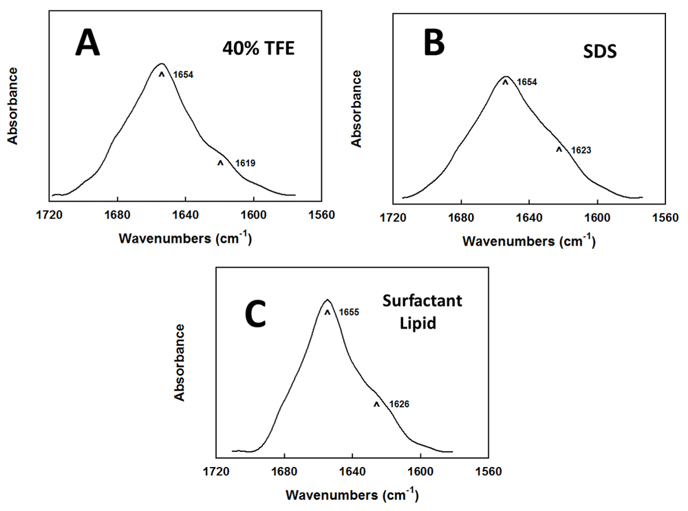
Attenuated total reflectance Fourier transform infrared (ATR-FTIR) spectral plots show the absorbance (in arbitrary units) as a function of wavenumbers (cm-1) (See Methods and Results). (A) Deuterated aqueous 40% TFE. (B) Deuterated aqueous SDS. (C) Deuterated aqueous surfactant lipid. In (A), (B) and (C), the IR spectra each show dominant α-helical components centered at 1654–1655 cm-1 (arrows), with minor bands at ~1619–1626 cm-1 (arrows) due to β-sheet, ~1682–1662 cm-1 due to loop-turn/bend, and ~1650–1637 cm-1 due to disordered or random conformations. Peptide concentrations were 470 μM for the TFE solvent spectra, and 10:1 lipid:peptide (mole:mole) for the SDS detergent and surfactant lipid spectra. The areas under each absorbance curve are normalized.
The secondary conformations for B-YL in lipid mimics (i.e., 40% TFE, aqueous SDS) or synthetic surfactant lipids [aqueous (DPPC:POPC:POPG 5:3:2 mole:mole:mole] were also studied with Circular Dichroism (CD) spectroscopy, to validate the above FTIR results. CD spectra for B-YL in these environments (Figure 3) were all similar, each indicating a major α-helical component characterized by a double minimum at 208 and 222 nm48–50. Deconvolutions of the Figure 3 spectra showed that B-YL is polymorphic, principally adopting α-helix (~45–52%), but with significant contributions (i.e., ~10–26% each) from loop-turn, disordered/random and β-sheet components (Table 1). Interestingly, the secondary conformation proportions determined from CD analysis for B-YL (i.e., α-helix > random ~loop-turn ~β-sheet) in both surfactant lipids and lipid mimetics are all compatible with B-YL folding as an α-helix hairpin17. Moreover, the overall maintenance of these secondary conformations from CD spectra in Table 1 additionally supports our FTIR findings that B-YL assumes a stable 3D-structure in environments of varying polarity.
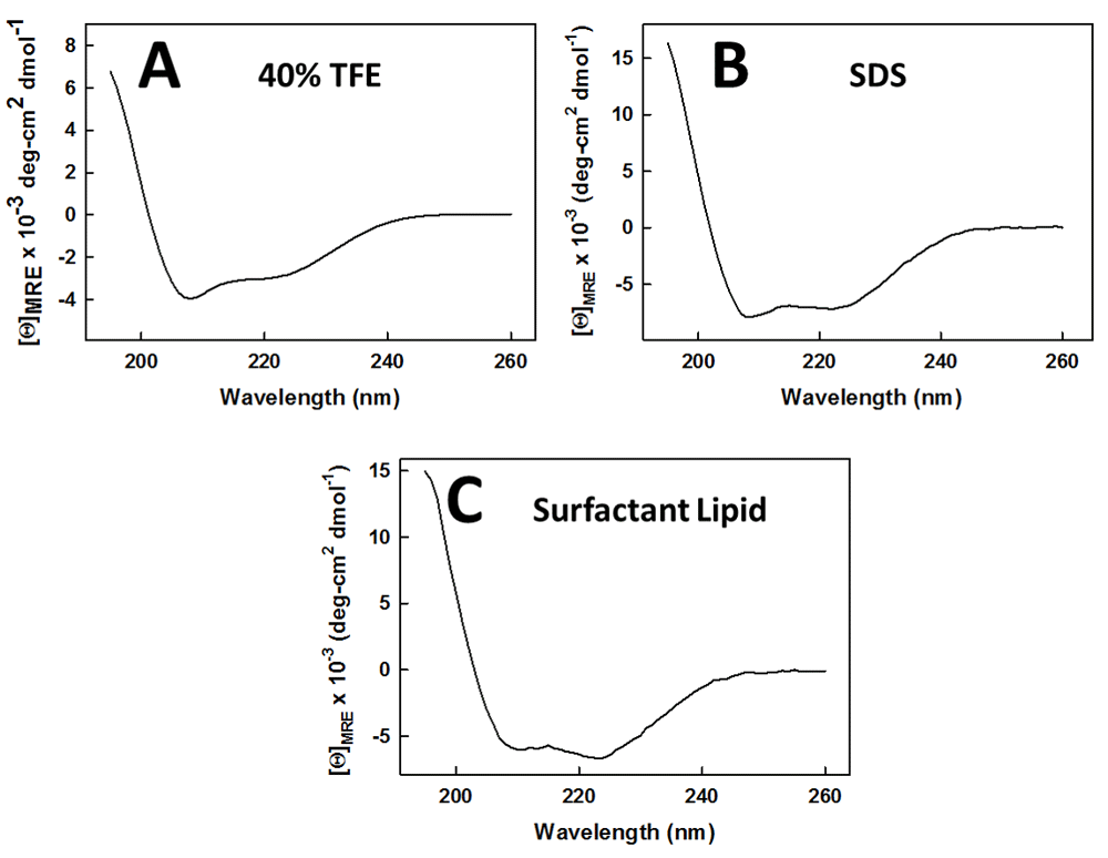
Circular Dichroism (CD) spectral plots show the mean residue ellipticities ([θ]MRE x 10-3 deg-cm2 dmol-1) as a function of wavelength (nm). (A) 40% TFE. (B) SDS. (C) Surfactant Lipid. The double minimum at 208 and 222 nm in each plot indicates that α-helix is the dominant secondary structure for B-YL in these environments. Peptide concentrations were 100 μM. The optical pathlength was 0.01 cm and the temperature was 37°C. Spectra represent the average of 8 scans.
Comparative FTIR spectroscopic studies were next performed on oxidized Super Mini-B (SMB) in lipid mimics and surfactant lipids to assess whether the B-YL substitutions in Figure 1 perturb the structure of the parent SMB. The secondary structures for SMB in lipid mimetics (i.e., deuterated 40 % TFE, deuterated aqueous SDS) and surfactant lipids (deuterated aqueous DPPC:POPC:POPG 5:3:2 mole:mole:mole) were investigated with conventional 12C-FTIR spectroscopy. Figure 4 shows that representative FTIR spectra of the amide I band for SMB in these environments were similar, each indicating a dominant α-helical component centered at ~1654–1655 cm-1 with a small low-field shoulder due to β-sheet at ~1618–1620 cm-134,47. Self-deconvolution of the FTIR spectra in Figure 4 demonstrated that SMB in either TFE, SDS or surfactant lipids folds with secondary structures that are characteristic of the α-helix hairpin (Table 1). Our finding that the respective secondary structure profiles for B-YL and SMB are similar in Table 1 suggests that the amino-acid substitutions in B-YL (e.g., four Cys residues replaced by Tyr) do not disrupt the α-helix hairpin conformation. Instead, these results raise the possibility that the replacement Tyr residues may stabilize the B-YL fold via a core of clustered tyrosines linking the N- and C-α-helices through noncovalent interactions involving aromatic rings (see below).
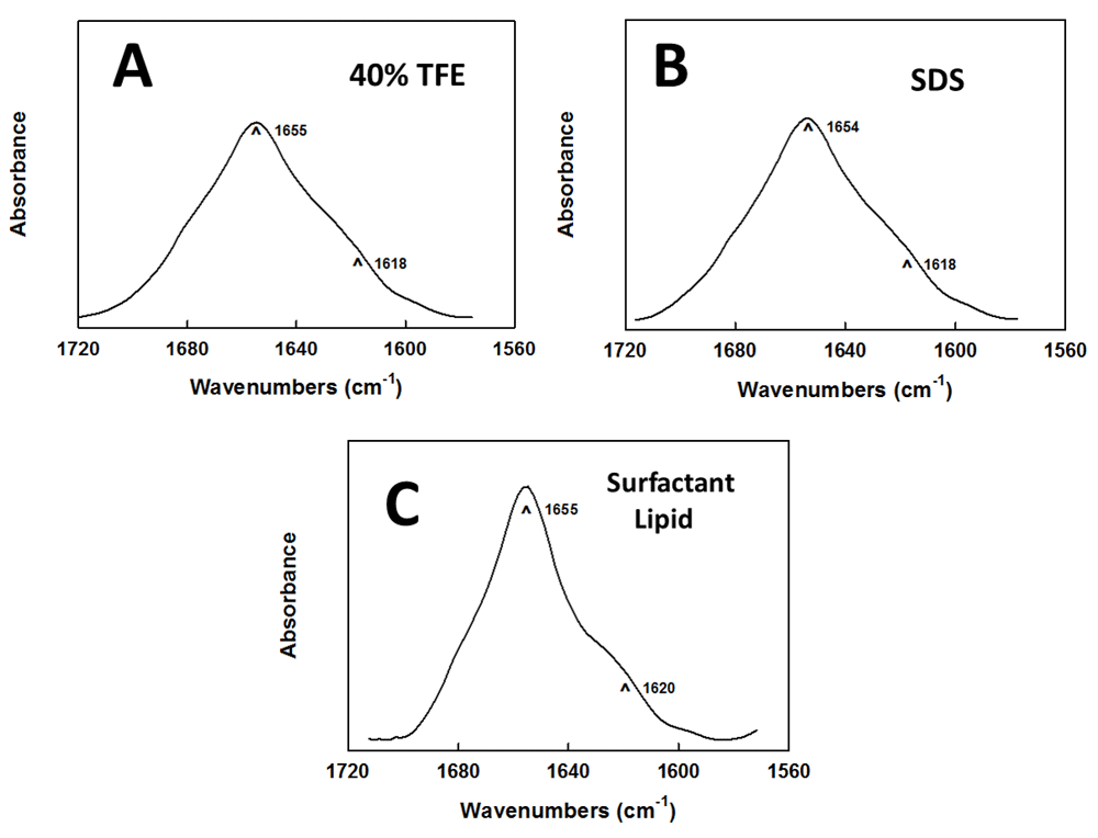
Attenuated total reflectance Fourier transform infrared (ATR-FTIR) spectral plots show the absorbance (in arbitrary units) as a function of wavenumbers (cm-1) (See Methods and Results ). (A) Deuterated aqueous 40% TFE. (B) Deuterated aqueous SDS. (C) Deuterated aqueous surfactant lipid. In (A), (B) and (C), the IR spectra each show dominant α-helical components centered at 1654–1655 cm-1 (arrows), with minor bands at ~1618–1620 cm-1 (arrows) due to β-sheet, ~1682–1662 cm-1 due to loop-turn/bend, and ~1650–1637 cm-1 due to disordered or random conformations. Peptide concentrations were 470 μM for the TFE solvent spectra, and 10:1 lipid:peptide (mole:mole) for the SDS detergent and surfactant lipid spectra. The areas under each absorbance curve are normalized.
MD simulations were subsequently conducted to obtain residue-specific information on B-YL in a surfactant lipid bilayer-water box. Although the above 12C-FTIR results on SP-B mimics (i.e., B-YL and SMB) are useful for experimentally assessing secondary structures averaged over the entire peptide, they cannot indicate the conformations of individual amino acids. With starting models based on homologous structures, however, MD runs in the Gromacs environment should provide our most accurate estimates of not only the 3D-conformations of SP-B mimics, but also the molecular topography of these peptides in the hydrated lipid bilayer. In the present work, MD simulations on B-YL were begun by first predicting the homologous 3D-structure by submitting the B-YL sequence (Figure 1B) to the I-TASSER web service. Three distinct models for B-YL were obtained from I-TASSER Version 5.1, and Model 1 with the highest C-score was selected (not shown). The accuracy of Model 1 was estimated from the following parameters: C-score of -0.73, TM-score of 0.62 ± 0.14 and RMSD of 3.6 ± 2.5 Å. C-score is a confidence score for evaluating the quality of I-TASSER models (between -5 to 2), with elevated values indicating a model with high confidence35,36. TM-score is a scale quantifying the similarity between two structures, with scores >0.50 signifying a model of correct topology and scores <0.17 implying random similarity35–37,51. Moreover, root mean square deviation (RMSD) is an average distance of all residue pairs in the two structures. The high C- and TM-scores, combined with the low RMSD, indicate that Model 1 will provide useful initial estimates of the secondary and tertiary structures for B-YL.
The resulting I-TASSER model for B-YL (not shown) predicts a C-terminal α-helix (residues P30-V37) connected to an N-terminal helix (residues Y8-L21) via a coil (residues I22-L29), which assumes a helix hairpin conformation52,53. The putative helix hairpin of B-YL executes a reverse turn after 8-residues with the I22-G25 component being the most prominent. Comparable to prior helix hairpins54, homology analysis of the B-YL sequence indicated high β-turn propensities for I22 to G25, allowing close interactions between the hydrophobic interfaces of the nearly antiparallel N- and C-helices. The helix hairpin fold of B-YL may be stabilized by a general increase in hydrophobicity due to Tyr and Leu substitutions (Figure 1A, B). Enhanced helix hairpin folding may also be due to the formation of clustered Tyr residues (i.e., Y7, Y8, Y11, Y34, and Y40) in the protein interior linking together the N- and C-helices through noncovalent interactions involving aromatic rings (not shown). The driving force behind this Tyr networking is at least partly due to “π-stacking” interactions of neighboring aromatic groups55,56, which were frequently observed in a survey of proteins deposited in the PDB57. Interestingly, Y7, Y8, and Y40 are oriented in a “pinwheel” arrangement with a 3-fold axis inside the protein core, similarly to how π-stacking interactions optimally organize benzene trimers in an early molecular mechanics study57. A second π-stacked configuration for the remaining Tyr residues was identified immediately adjacent to the pinwheel trimer, in which Y34 and Y11 nearly adopt a 1p dimer configuration with their tyrosine rings off-centered and parallel displaced55,57. The I-TASSER model further forecasts a flexible coil for the N-terminal insertion sequence (F1-P6), which permits this hydrophobic segment to interact with the π-stacked tyrosine residues within the B-YL interior. Lastly, the space-filling I-TASSER model indicates that the folding of B-YL into a helix hairpin produces a compact globular protein, exhibiting a core of hydrophobic residues and a surface of aqueous-exposed and polar residues (not shown).
MD simulations of B-YL were next calculated by porting the above I-TASSER Model 1 into aqueous DPPC:POPC:POPG bilayers with potassium counterions to maintain electrical neutrality. This lipid mixture was chosen for in silico studies because it optimizes the surfactant activity of our SP-B mimics in both in vitro and in vivo assays. Simulations were then carried out for 0–500 nsec using the Charmm 36 implementation for lipids in the Gromacs (Version 5.1.1) environment40. The representative “500 nsec” structure for B-YL in lipids (Figure 5A) largely agrees with the I-TASSER model on which it is based. Similar to the original I-TASSER model, Figure 5A indicates that the “500 nsec” model in surfactant lipids is folded as a helix-hairpin-helix, in which an N-terminal α-helix (residues R12-L21) connects to a C-terminal α-helix (P30-L36) via a turn-loop (I22-L29). The high β-turn propensity of B-YL in Figure 5A (i.e., P23 – G26; green wire sidechains) allows close interaction between the hydrophobic interfaces of the nearly antiparallel N- and C-helices. The 500 ns B-YL model also predicts flexible coils for the N-terminal insertion (F1 – Y11) and the C-terminal (V37 – S41) sequences. Lastly, space-filling I-TASSER and 500 ns MD simulation models for B-YL each produced a compact globular protein, exhibiting a core of hydrophobic residues and a surface of aqueous-exposed and polar residues (not shown).
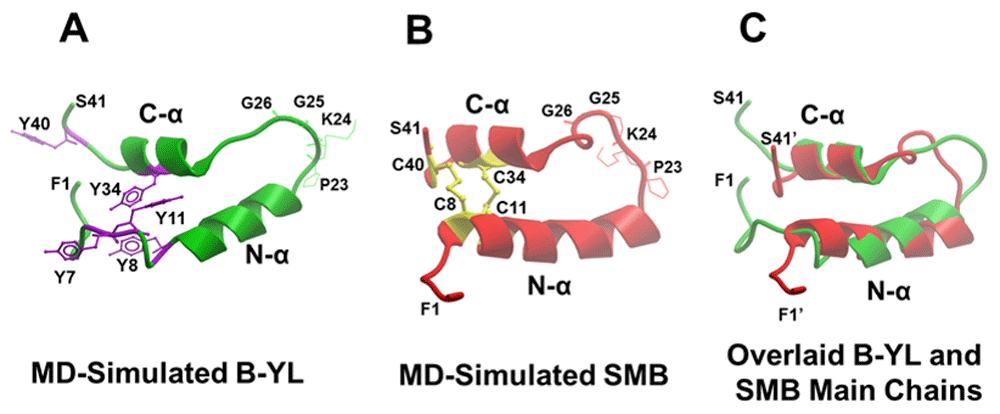
Molecular Dynamics (MD) simulations were carried out on the Super Mini-B mimic (B-YL) and oxidized Super Mini-B (SMB) (see Methods and Results). Main-chain folding of peptides is displayed in ribbon format, with lipids and water omitted for clarity. (A) The green ribbon MD model for B-YL is shown, in which Tyr replaces Cys at residues 8, 11, 34, and 40 of the parent SMB with purple stick sidechains (see Figure 1). This 500 nsec simulation predicts a helix-hairpin, in which an N-terminal α-helix (i.e., green N-α, residues R12 – L21) connects to a C-terminal α-helix (i.e., green C-α, residues P30 – L36) via a turn (green residues I22 – L29). The B-YL model also predicts flexible coils for the N-terminal insertion (F1 – Y11) and the C-terminal (V37 – S41) sequences. The B-YL fold is stabilized by a hydrophobic core of clustered Tyr (Y8, Y11, and Y34) linking together the N- and C-helices through noncovalent interactions involving aromatic rings. (B) The ribbon model for SMB at 490 nsec forecasts a helix-hairpin conformation, in which an N-terminal α-helix (i.e., red N-α, residues Y7 – M21) connects to a C-terminal α-helix (i.e., red C-α, residues P30 – L36) via a turn (red residues I22 – L29). The SMB model also predicts flexible coils for the N-terminal insertion (F1 – P6) and the C-terminal (V37 – S41) sequences. SMB is oxidized with covalent disulfides (i.e., C8–C40, and C11–C34; yellow stick and ribbon figures) lying deep inside the hydrophobic core. (C) The ability of the B-YL peptide to mimic the helix-hairpin folding of its parent SMB was tested with a superimposition of the ribbon backbone of SMB (red) with that of B-YL (green).
Although major changes were not observed between the I-TASSER and 500-nsec models for B-YL, there were nevertheless significant minor differences which we attribute to the 500 nsec-equilibration of the I-TASSER structure in the aqueous lipid-bilayer box. For example, note that the N α-helix of the “500 nsec” model is shorter by four residues, with Y8, W9, L10, and Y11 not participating in the N α-helix. Concurrently with the fraying of the N-helix by one turn, there is a reorganization of the five tyrosine residues in the “500 nsec” model. Namely, the pinwheel trimer and 1p dimer in the I-TASSER B-YL model convert to a distorted pinwheel trimer (i.e., Y8, Y11 and Y34) in the hydrophobic interior of the 500 nsec B-YL, with Y7 and Y40 migrating to the polar surface (Figure 5A). The approximate 3-fold axis relating the three tyrosines is seen more clearly by reorienting the 500 nsec model so that the symmetry axis is perpendicular to the plane of the paper (see “X"), with the Tyr representations in purple stick (Figure 6A) and space-filling (Figure 6B), respectively. This three-fold axis is designated an “approximate” symmetry axis because it is strictly valid for all three tyrosine ring structures, but not for the hydroxyl of Y8, which faces an opposite direction than those of the hydroxyls for either Y11 or Y34 (Figure 6A, B). Figure 6 confirms that the 500 nsec model B-YL folds as a helix hairpin, with the approximate pinwheel trimer (Y8, Y11, and Y34) bridging together the N- and C-α helices through non-covalent interactions. The space-filling 500 nsec model for B-YL further demonstrates a globular protein, in which the pinwheel Tyr trimer embeds with other hydrophobic residues in the interior, while hydrophilic groups face the exterior (not shown). Notably, the pinwheel Tyr trimer in Figure 6 may act as a hydrophobic core around which other hydrophobic residues assemble, but from which all water is absent. Because water disrupts secondary structure by attacking amide-amide hydrogen bonds, the stability of the B-YL conformation for 500 nsec in Figure 5A and Figure 6 may be due not only to non-covalent interactions between hydrophobic residues (e.g., π-stacking of Tyr residues) but also to water exclusion from the hydrophobic interior (i.e., solvophobic forces)55,56.
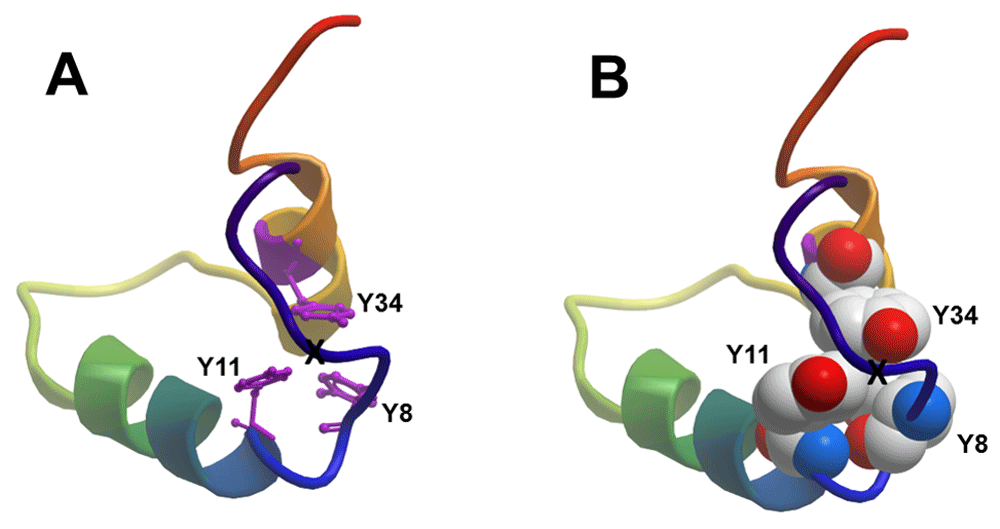
Graphical modeling and MD simulations were carried out as described in Figure 5A and Methods. Main-chain folding of 500 nsec B-YL is shown in the ribbon-rainbow format, with the N-terminal insertion sequence in blue, the N-terminal helix in blue-green, the turn hairpin in green yellow and the C-terminal helix in yellow-orange. Three of the tyrosine residues (i.e., Y8, Y11, and Y34) are organized as a distorted pinwheel trimer in the hydrophobic interior of the 500 nsec B-YL that links together the N- and C-helices through non-covalent interactions. An approximate 3-fold axis relating the three tyrosines in Figure 5A is seen more clearly by rotating the 500 nsec model so that the symmetry axis is perpendicular to the plane of the paper (see “X”). This three-fold axis is designated an “approximate” symmetry axis because it is strictly valid for all three tyrosine ring structures, but not for the hydroxyl of Y8, which faces an opposite direction than those of the hydroxyls for either Y11 or Y34. (A) Graphical representation of the rotated 500 nsec B-YL model is in ribbon-rainbow format, with tyrosine sidechains as purple sticks. (B) Graphical representation of the rotated 500 nsec B-YL model is in ribbon-rainbow format, with tyrosine sidechains as space-filling (CPK).
The topological organization of B-YL in the membrane bilayer may also be characterized using the above all-atom (500 nsec) MD simulation of B-YL in hydrated lipid surfactant (DPPC:POPC:POPG 5:3:2 mole:mole:mole) at 37°C. Figure 7A shows a cross-sectional view of the ribbon model for the 500 nsec B-YL in the lipid bilayer. Here, the buried Y8, Y11 and Y34 tyrosines (i.e., purple stick sidechains) bridge the N- and C-α helices through noncovalent aromatic interactions, while the surface-exposed Y7 and Y40 interact with water molecules or polar lipid headgroups (see also Figure 5A). The axes for the N- and C-α helices are each nearly parallel to the bilayer plane. However, the N-α helix lies deeper in the bilayer subjacent to the lipid headgroup, while the C-α helix binds to the more polar lipid-water interface (Figure 7A). This differential partitioning may be just due to differences in the hydrophobicities of the two helices because MPEx analysis41 indicated a higher hydropathy (i.e., more hydrophobic) for the N-α-helix than for the C-α helix (i.e., 4.92 vs. -0.02 kcal/mol, respectively). The 500 nsec – MD simulation model for B-YL in aqueous surfactant lipids also demonstrated the absence of any water or lipid within its hydrophobic core (not shown). The systematic exclusion of water from the protein interior mediated by the three “π-stacked” tyrosine residues may account for why B-YL folds as a helix-hairpin for simulation times (t) =500 nsec.
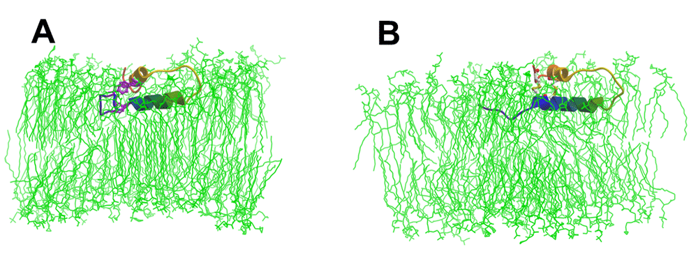
Graphical modeling and MD simulations were carried out as described in Figure 5 and Methods. Main-chain folding for peptides is shown in the ribbon-rainbow format, with the N-terminal insertion sequence in blue, the N-terminal helix in blue-green, the turn hairpin region in green-yellow and the C-terminal helix in yellow-orange. Lipids are shown as green stick figures, while water is left out for clarity. (A) The ribbon model for the 500 nsec B-YL conformer folds as a helix-hairpin, with the Y8, Y11 and Y34 tyrosine residues (i.e., purple stick figures) bridging the N- and C-α helices through noncovalent aromatic interactions. The axes for the N- and C-α helices are each nearly parallel to the bilayer plane. However, the N-α helix lies deeper in the bilayer subjacent to the lipid headgroup, while the C-α helix binds to the more polar lipid-water interface. (B) The ribbon model for the 499 nsec SMB conformer also adopts a helix-hairpin structure, with the disulfide bonds (i.e., two yellow stick figures) covalently linking the N- and C-α helices. The respective topographical organizations for SMB and B-YL are both similar. Namely, the axes for the N- and C-α helices of SMB are each nearly parallel to the bilayer plane. Moreover, the N-α helix of SMB lies deeper in the bilayer beneath the lipid headgroup, while the corresponding C-α helix binds to the more polar lipid-water interface.
Importantly, partial validation of the 500 nsec MD structure for B-YL in aqueous surfactant lipids (Figure 5A) is provided by our above spectroscopic findings. The secondary structures obtained from either FTIR or CD spectra of B-YL in surfactant lipids (Figures 2C and Figure 3C; Table 1) are generally in good agreement with those predicted in the 500 nsec model (Figure 5A). For example, Figure 8A indicates that the respective proportions of secondary structures for B-YL obtained from FTIR self-deconvolutions are compatible with those determined using MD simulations, with the experimental and theoretical techniques each showing high α-helix and smaller contributions due to loop-turn, disordered and β-sheet. These results indicate experimental confirmation of critical features for our MD model of B-YL folding as an α-helix-hairpin (Figure 5A).
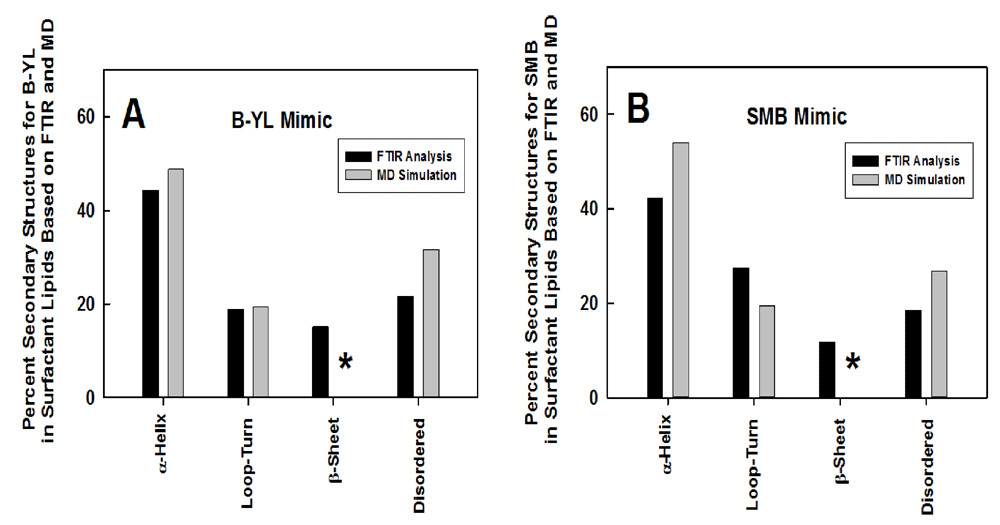
The SMB mimic is oxidized Super Mini-B with covalent disulfide bonds at C8–C40 and C11–C34, while the B-YL mimic lacks disulfide linkages with Tyr substituted for Cys at residues 8, 11, 34 and 40 and also Leu replacing Met at residues 21 and 28 (see Figure 1). (A) Plot of % conformations assessed from FTIR spectra (black bars) of the B-YL mimic in DPPC:POPC: POPG (5:3:2 mole:mole:mole) bilayers (Table 1) vs. the corresponding structures calculated from DSSP analysis (i.e., H-bond estimation algorithm) of an MD simulation of B-YL (gray bars) in an aqueous DPPC:POPC:POPG lipid box for 500 nsec. The respective proportions of secondary structures for B-YL obtained from FTIR deconvolution are in good agreement with those determined using MD simulations, with each technique showing α-helix > Loop-Turn ~ Disordered = β-sheet. The absence of any ≥-sheet in our MD model is shown by the asterisk (*). (B) Plot of % conformations assessed from ATR-FTIR spectra (black bars) of the SMB mimic in DPPC:POPC: POPG (5:3:2 mole:mole:mole) bilayers (see Table 1) vs. the corresponding structures calculated from DSSP analysis of an MD simulation of SMB (gray bars) in an aqueous DPPC:POPC:POPG lipid box for 490 nsec. The respective proportions of secondary structures for SMB obtained from FTIR spectral deconvolution are comparable to those determined with MD simulations, with each technique showing α-helix > Loop-Turn ~ Disordered = β-sheet. The absence of any β-sheet in our MD model is shown by the asterisk (*).
MD simulations were likewise performed on oxidized Super Mini-B (SMB) in the surfactant lipid bilayer, to more directly assess whether the amino-acid replacements in B-YL (i.e., four Cys residues substituted with Tyr and two Met residues with Leu) significantly perturbed the α-helix hairpin conformation or its insertion in the hydrated lipid bilayer. As with B-YL, MD simulations on SMB were started by first predicting the homologous 3D-structure by submitting the reduced SMB sequence (Figure 1A, but without the disulfide bonds) to the I-TASSER web service (see Methods). Four separate models for reduced SMB were obtained from I-TASSER Version 5.1, and Model 1 with the highest C-score was selected for further analysis. Model 1 was estimated to be the most accurate of the four models using the following parameters: C-score of 0.01, TM-score of 0.71 ± 0.11 and RMSD of 2.3 ± 1.8 Å (see above and Methods)35–37,51. The I-TASSER Model 1 for the reduced SMB predicts a C-terminal α-helix (residues P30-L36) connected to an N-terminal helix (residues C8-M21) via a loop-turn (residues I22-R27), which adopts a helix hairpin conformation. Because the distances between two sulfur atoms in the C8-C40 and C11-C34 pairings are only 3.5 and 5.0 Å, respectively, Hyperchem 8.0 readily forms oxidized SMB by converting the four cysteine residues in reduced SMB to the known disulfide bonds.
The above oxidized SMB was then ported into the hydrated surfactant lipid bilayer, and MD simulations were run for 0–500 nsec, permitting detailed structural comparisons between the parent SMB and its daughter peptide B-YL. Figure 5B shows a representative “490 nsec” structure for SMB folding as a α-helix hairpin, with an N-terminal α-helix (i.e., red N-α, residues Y7 – M21) connecting to a C-terminal α-helix (i.e., residues P30 – L36) via a turn (i.e., residues I22 – L29). Experimental support for this helix-hairpin model comes from 12C-FTIR spectroscopy, in which self-deconvolutions obtained from spectra of SMB in lipid surfactant indicated high α-helix and smaller components due to loop-turn, disordered and β-sheet (Figure 4C and Figure 8B; Table 1)17. The helix-hairpin for the 490 nsec model (Figure 5B) produces a reverse turn after 8-residues with the P23-G26 component (red wire sidechains) being the most prominent. Similar to earlier helix-hairpins, the 490 nsec model indicated high β-turn propensities for I22 to G25, allowing close interactions between the hydrophobic interfaces of the nearly antiparallel N- and C-helices. The oxidized SMB additionally predicts flexible coils for the N-terminal insertion (F1 – P6) and the C-terminal (V37 – S41) sequences, as well as covalent disulfides (i.e., C8–C40 and C11–C34) deep within the hydrophobic core that bridge together the N – and C – α-helices. The space-filling 490 nsec model for SMB (not shown) additionally indicates a globular protein, in which the hydrophobic disulfide bonds embed with other hydrophobic residues in the interior to quantitatively exclude water, while hydrophilic groups face the exterior. Given that water perturbs secondary structures by attacking amide H-bonds, the stability of the SMB conformation for 490 nsec in Figure 5B may be due not only to covalent disulfide bonds and non-covalent interactions between hydrophobic residues but also to water exclusion from the protein interior (e.g., solvophobic forces)17,55,56. Lastly, the insertion of SMB into the lipid bilayer was assessed using an all-atom MD simulation (i.e., a 499 nsec conformer) of SMB in the hydrated lipid surfactant at 37°C. Figure 7B indicates a cross-sectional view of the ribbon model for the 499 nsec SMB in the lipid bilayer, in which the axes for the N- and C-α-helices are each nearly parallel to the bilayer plane. Nevertheless, the N- α helix lies more buried in the bilayer underlying the lipid headgroup, while the C- α helix binds to the more polar lipid-water interface (Figure 7B). MPEx analysis of SMB indicated higher hydropathy (i.e., more hydrophobic) for the N- α-helix than for the C- α-helix (i.e., 3.55 vs. 1.87 kcal/mol, respectively)41, and this may be responsible for the deeper penetration of the N- α-helix into the bilayer.
Head-to-head comparisons were next conducted on MD simulated models of B-YL and its parent SMB in hydrated lipid surfactant bilayers at ~500 nsec, to determine whether the amino-acid replacements in B-YL significantly perturbed the α-helix hairpin conformation or its insertion in the lipid bilayer. The 500 nsec model of B-YL in Figure 5A is very similar to the corresponding 490 nsec model of the parent SMB in Figure 5B, except for the fraying of the N-terminus of the N-α helix (residues 8-11; one turn), and also the substitution of a distorted tyrosine pinwheel (i.e., purple Y8, Y11, and Y34) that draws together the C-α and N-α helices in Figure 5A instead of the two disulfide linkages (i.e., yellow C8–C40 and C11–C34) in Figure 5B. The fraying of the N-α helix occurs concurrently with the formation of the distorted tyrosine pinwheel in B-YL, suggesting that an unwinding of one helical turn is energetically required to accommodate the tyrosine reorganization of B-YL in Figure 5A. Interestingly, the location of the distorted tyrosine pinwheel in B-YL (Figure 5A and Figure 6) overlaps that of the C11–C34 disulfide bond in SMB (Figure 5B), suggesting that the tyrosine trimer is well positioned to promote the α-helix hairpin conformation in B-YL (see above and Figure 6). The ability of the B-YL peptide to mimic the helix-hairpin folding of its parent SMB was further tested with the automated multiple structural superposition tools in the MolBrowserPro environment. Figure 5C shows a weighted iterative superposition that was performed with the visible atoms of the aligned residues, using SMB as the static object. The output of the superimposition in Figure 5C is shown with the ribbon backbone of SMB (red) overlaid with that of B-YL (green). There is good overlap between the two structures, with the best seen in the stable N- and C-helical cores and less agreement between the more flexible turn and N- and C-terminal regions. These results indicate that the tyrosine substitutions create a B-YL daughter peptide that successfully mimics essential helix-hairpin features of the parent SMB. Because high in vitro and in vivo surfactant activities are associated with SMB adopting an α-helix hairpin conformation10,14,17, it will be worthwhile to investigate similar functional correlates with B-YL.
Another direct comparison was performed on MD simulated models of B-YL and SMB in hydrated lipid surfactant bilayers, to assess if the amino-acid substitutions in B-YL disturbed its topological organization in the lipid bilayer (Figure 7A, B). As noted above, Figure 7B shows a cross-sectional view of 499 nsec SMB in the lipid bilayer, in which the N- and C-α-helices are each nearly parallel to the bilayer plane, but the N-α helix lies deeper in the bilayer than the disulfide-linked C-α helix. The corresponding view of 500 nsec B-YL (Figure 7A) demonstrates insertion of its α-helix hairpin into the lipid bilayer which is remarkably analogous to that of 499 nsec SMB (Figure 7B). Accordingly, B-YL and SMB may be each classified as a tightly bound peripheral membrane protein, which interacts with superficial portions of the phospholipid bilayer and lacks transmembrane segments that span the width of the bilayer. The finding of similar topological organizations for B-YL and SMB in the lipid bilayer provides further support for the hypothesis that B-YL may likewise mimic the surfactant activities of SMB (see below).
Captive bubble surfactometry of B-YL and SMB surfactants (3% peptide in DPPC:POPC:POPG 5:3:2 mole:mole:mole), Curosurf®, and surfactant lipids alone demonstrated excellent surface activity of B-YL (Figure 9). Surface activity of BYL and SMB surfactant and Curosurf® were consistently and equally low during quasi-static cycling with values ≤1 mN/m, indicating that the modifications in the B-YL peptide did not lead to a loss in in vitro surface activity compared to its parent peptide SMB. In contrast, minimum surface tension values of surfactant lipids alone far exceeded those of B-YL, SMB and Curosurf® surfactant and amounted to ~18 mN/m (p<0.001).
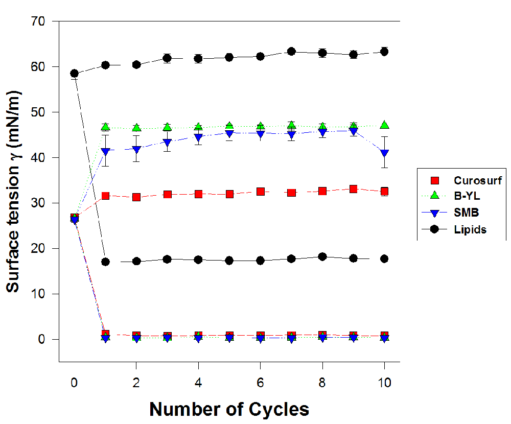
Surface activity of 3% B-YL and oxidized Super Mini-B (SMB) in surfactant lipids were compared with a clinical surfactant (Curosurf®) as positive control and surfactant lipids alone (DPPC:POPC:POPG 5:3:2 mole:mole:mole) as negative control. The lower part of each curve indicates minimum surface tension and the upper part indicates maximum surface tension during 10 compression-expansion cycles. Minimum surface tension values of B-YL and SMB surfactant were similar to those of Curosurf®. Values are mean ± SEM of N=4-5.
Animal experiments directly examined the in vivo pulmonary activity of B-YL surfactant when instilled intratracheally in ventilated young adult rabbits with surfactant deficiency and impaired lung function induced by repeated saline lung lavages (Figure 10). Surfactant was administered by intratracheal instillation after the PaO2 was reduced to stable levels <100 mmHg, which is far below the threshold clinical criteria for ARDS of a PaO2 <200 mmHg and for ALI of <300 mmHg when breathing 100% oxygen. For this timeframe of study, this model reflects a relatively pure state of surfactant deficiency in animals with mature lungs. Rabbits receiving B-YL, SMB and Curosurf® surfactant had significantly improved arterial oxygenation over the 2 h period of study post-instillation compared to control rabbits instilled with surfactant lipids alone (Figure 10, p<0.001). Dynamic lung compliance also significantly improved over the same period of post-instillation study in rabbits treated with B-YL and SMB surfactants and Curosurf® compared to lipid-only controls (Figure 10). The differences between B-YL and SMB surfactants and Curosurf® were not statistically significant, indicating that the modifications in the SP-B peptide mimic B-YL did not lead to a loss in in vivo surface activity.
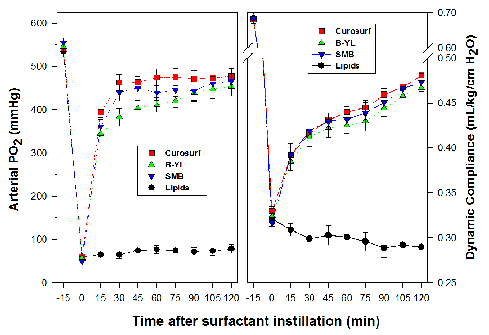
Oxygenation (arterial PO2 in mmHg) and dynamic lung compliance (mL/kg/cm H2O) of 3% B-YL (n=9) and oxidized SMB (n=8) in surfactant lipids were compared with a clinical surfactant (Curosurf®) (n=8) or lipids alone (DPPC:POPC:POPG 5:3:2 mole:mole:mole) (n=8). Surfactant (100 mg/kg) was administered intratracheally as a bolus at time 0. Values are mean ± SEM. Differences in oxygenation and dynamic compliance between B-YL, SMB and Curosurf surfactants were not statistically significant, but differences with lipids alone were significant (p<0.001).
The basic premise tested here was whether the ‘sulfur-free’ and ‘oxidation-resistant’ B-YL peptide, a 41-residue Super Mini-B (SMB) variant that has its four cysteine residues replaced by tyrosine and its two methionine residues replaced by leucine, would fold with the same α-helix hairpin conformation earlier shown by SMB to correlate with high in vitro and in vivo surfactant activities8,10,14,17.
Circular dichroism (CD) and FTIR spectroscopy of ‘B-YL’ in surfactant lipids showed secondary structures compatible with the peptide folding as an α-helix hairpin, similar to that of SMB in lipids. All-atom (500 nsec) MD simulations confirmed that B-YL maintained its α-helix hairpin in a lipid bilayer, matching the hairpin obtained from the corresponding MD simulation of SMB. In contrast to the disulfide-reinforced helix-turn of SMB, however, the B-YL fold was stabilized by a core of clustered Tyr linking the N- and C-α-helices through noncovalent interactions involving “π-stacked” aromatic rings. MD simulations also indicated similar topological organizations for B-YL and SMB in the hydrated bilayers. Namely, the axes for the N- and C-α-helices of both peptides were nearly parallel to the membrane plane, and also the N-α-helix for both lies deeper in the bilayer subjacent to the lipid headgroup, while the C-α-helix binds to the more polar lipid-water interface. Captive bubble surfactometry indicated excellent surface activity for B-YL surfactant, while also showing good oxygenation and dynamic compliance in lavaged, surfactant-deficient adult rabbits, an animal model of acute lung injury (ALI) and acute respiratory distress syndrome (ARDS).
Our ‘sulfur-free’ and ‘oxidation-resistant’ B-YL mimic may therefore prove to be superior to its parent (oxidized) SMB for treating ALI/ARDS patients for several reasons. First, B-YL is less expensive and faster to synthesize than SMB because it omits an oxidation step and is self-folding. Second, the substitution of Cys and Met residues with Tyr and Leu, respectively, may render the B-YL mimic less susceptible to the ROS inactivation typically observed in ALI/ARDS patients.
Lung immaturity and surfactant deficiency are the main cause of respiratory distress syndrome (RDS) in very preterm infants. Mechanical ventilation and exposure to high oxygen concentrations may lead to an inflammatory lung process resulting in bronchopulmonary dysplasia (BPD), a chronic lung disease of preterm infants. Use of antenatal steroids, administration of exogenous surfactant, and advanced modes of ventilation have shown only limited benefits in preventing RDS and BPD. In a neonatal rat model of hyperoxia-induced lung injury, we found that nebulized PPARγ agonist pioglitazone (PGZ) with B-YL surfactant accelerates lung maturation and prevents neonatal hyperoxia-induced lung injury more than with either modality alone, thereby potentially preventing BPD more effectively58. These findings suggest a potential role for B-YL surfactant as a vehicle for intrapulmonary drug therapy.
The ‘sulfur-free’ and ‘oxidation-resistant’ B-YL forms an amphipathic helix-hairpin in surfactant liposomes with high surface activity, and is functionally similar to its parent (oxidized SMB) and native SP-B. B-YL’s resistance against free oxygen radical damage provides an extra edge over oxidized SMB in the treatment of respiratory failure in newborn infants with RDS and children and adults with ALI/ARDS.
Raw data are available on OSF: http://doi.org/10.17605/OSF.IO/6295P59
Data are available under the terms of the Creative Commons Zero "No rights reserved" data waiver (CC0 1.0 Public domain dedication).
| Views | Downloads | |
|---|---|---|
| Gates Open Research | - | - |
|
PubMed Central
Data from PMC are received and updated monthly.
|
- | - |
Provide sufficient details of any financial or non-financial competing interests to enable users to assess whether your comments might lead a reasonable person to question your impartiality. Consider the following examples, but note that this is not an exhaustive list:
Sign up for content alerts and receive a weekly or monthly email with all newly published articles
Register with Gates Open Research
Already registered? Sign in
If you are a previous or current Gates grant holder, sign up for information about developments, publishing and publications from Gates Open Research.
We'll keep you updated on any major new updates to Gates Open Research
The email address should be the one you originally registered with F1000.
You registered with F1000 via Google, so we cannot reset your password.
To sign in, please click here.
If you still need help with your Google account password, please click here.
You registered with F1000 via Facebook, so we cannot reset your password.
To sign in, please click here.
If you still need help with your Facebook account password, please click here.
If your email address is registered with us, we will email you instructions to reset your password.
If you think you should have received this email but it has not arrived, please check your spam filters and/or contact for further assistance.
Comments on this article Comments (0)