Keywords
biosafety, laboratory biocontainment, bacteria, risk assessment, flow cytometer
biosafety, laboratory biocontainment, bacteria, risk assessment, flow cytometer
Flow cytometry techniques have been used to analyse bacteria for several decades1,2, and for assessing the effects of antimicrobial agents since the 1980s3–5. The bacteria analysed then included species that can be safely handled on an open bench in a suitably equipped microbiology laboratory, providing a series of standard biosafety procedures are adhered to6. Concerns about laboratory biosafety and containment increased after 20017,8 and led to higher physical containment levels for select biothreat agents and some bacterial species prone to transmission by aerosols generated during laboratory procedures such as Neisseria meningitidis9,10. Given these concerns about bioaerosol transmission risks, it is not surprising that standards for bioaerosol risk assessment and mitigation have been recommended for flow cytometry-assisted cell sorting protocols11,12. Our use of flow cytometry was not for cell sorting, but for a less aerosol-prone cellular analysis. We commenced use of our analytic flow cytometer in a physical containment two (PC2) laboratory while developing a flow cytometry-assisted antimicrobial susceptibility test (FAST) assay method with Klebsiella pneumoniae13. Though the cytometer we used had no cell sorting function, we decided to conduct an assessment of its ability to generate bacterial aerosols before progressing with any analysis of potentially more hazardous aerosol-transmitted species such as Neisseria meningitidis, Mycobacterium tuberculosis and Burkholderia pseudomallei.
This study aimed to detect airborne bacteria during operation of an acoustic-enhanced flow cytometer (AFC) (Attune NxT, Thermo Fisher Scientific, Eugene, OR) and to compare the detected bacteria with those cultured from air samples collected in the same laboratory space when the cytometer was not in use, and in a diagnostic clinical microbiology laboratory.
Laboratory locations. Two laboratory locations in adjacent buildings were compared. One was a university research laboratory equipped with air conditioning and high efficiency purified air (HEPA) filtration, two AFCs, and two class II biosafety cabinets, peripheral benches and one central bench. The other was a large, open plan clinical diagnostic microbiology laboratory operating a range of high throughput bacteriology procedures serving an on-campus 700 bed teaching hospital and an extensive regional hospital network.
Workflow. The air sampling study was conducted during a one month period during which the research laboratory was intensively used by up to nine people at one time during office hours, often with both flow cytometers in use at once. Each flow cytometer procedure was staffed by one cytometer operator and another scientist preparing bacterial suspensions for FAST and other cytometer assays, plus at least one of the above authors engaged in the air sampling procedure. The majority of flow cytometer use analysed the K. pneumoniae isolates previously reported13. The other two species in use were Streptococcus pneumoniae and Burkholderia thailandensis. The clinical laboratory was staffed between 7.30am and 9pm by up to 20 people, with 1–3 people per side of each laboratory bench, conducting predominantly manual procedures with liquid and solid bacterial cultures. While clinical specimens were opened and blood cultures were subcultured in a class 2 biosafety cabinet, the majority of bacteriologic procedures were performed on the open bench in accordance with clinical laboratory PC2 safety policy6.
Air sampling. Air sampling was performed with a sieve impaction air sampler (MAS-100 Eco, EMD Millipore, Merck) that drew a defined volume of room air over an agar plate positioned in the air sampling unit under the air-permeable lid. The sampling rate was set at 100 L per minute for 10 minutes. The lid surfaces were cleaned with isopropyl alcohol before and after each use in accordance with the manufacturer’s instructions. Two types of media were used; 5% horse blood agar (HBA) and MacConkey agar (MAC). Air was sampled for identical periods, volumes and locations to produce a pair of samples on each occasion. Air sampling was conducted before, during and at the end of a day to give a range of times reflecting different levels of research laboratory use and occupation. The same pattern of sampling was conducted for comparison in the clinical bacteriology laboratory. Two sampling locations were used in the research laboratory. The first was immediately adjacent to the sample introduction port of the AFC between the cytometer and its operator. For comparison, the second sampling site was on the preparation bench behind the operator approximately 2.5 m from the flow cytometer. These two locations did not apply in the clinical laboratory where there was no flow cytometer. Instead, air sampling was conducted in one location at the open bench where wound or urine swabs were plated directly onto agar, and adjacent to the rotary plating device used to inoculate agar plates with bacterial suspensions for disk diffusion antimicrobial susceptibility tests. Further sets of air samples were collected on multiple occasions after a detergent and ethyl alcohol clean of all research laboratory surfaces, and replacement of laboratory gowns. The cleaning procedures in the clinical laboratory comprised ethyl alcohol wipe down of laboratory bench and external equipment surfaces at the end of each shift, but no regular cleaning of storage shelves and other horizontal surfaces. These practices were not changed during the course of this study.
Equipment and bench surfaces. On completion of repeated air sampling over one month, surface swabs were collected from the external flow cytometer housing above, below and around the flow sample introduction port, the nearby open bench and the preparation bench opposite before any cleaning had occurred. Cotton swabs were immersed in sterile 0.9% normal saline before rubbing vigorously over the test surface, and then used to inoculate FBA and MAC. Duplicate swabs were collected at each site to avoid loss of bacteria to the first medium inoculated.
Bacterial counts. Colony forming units on both types of solid media were recorded after 24 hr incubation at 37°C and expressed as CFU/1000 L air, and CFU/plate for swabs..
Bacterial identification. All bacteria growing on MAC were identified using the clinical bacteriology laboratory’s identification protocol. In short, after macroscopic examination and discretionary Gram stain analysis, definitive identification is by matrix-assisted laser desorption ionisation time of flight mass spectrometry (MALDI-TOF) from an extended mass spectrometer profile (MSP) library. Initial borderline identification is repeated on the same sample on the stainless steel target with repeated MALDI-TOF analysis. Potential clinically significant isolates known to be problematic by MALDI-TOF or showing borderline acceptable identification were then subject to supplementary methods such as substrate utilisation panels (e.g. API20E, BioMerieux, France). For bacteria not growing on MAC but present on BA, the commonest colony types were identified plus any that resembled the three bacteria being interrogated by flow cytometry. A limit was set at the six most common colony types because this was the maximum distinguishable on an 8cm diameter agar plate when using a standard dilutional streaking method for swab samples.
Statistical analysis. Column statistics, Chi squared test and non-parametric tests (Mann-Whitney U test) were conducted with Prism statistical software (Prism 6.0, GraphPad, San Diego, CA).
The bacteria isolated from over 50,000 L of air sampled in the research laboratory before, during and after flow cytometer operation did not yield a single cultured colony of K. pneumoniae, S. pneumoniae or B. thailandensis (Table 1A, Table 1B). None of these species was recovered from either medium during the air sampling period, nor were they recovered from surface swabs taken above, below and around the flow cytometer sample introduction port or other research laboratory surfaces. Bacterial suspensions grown on HBA for experiments conducted on the flow cytometer remained culturable throughout the air sampling period. The bacteria we isolated from air samples in both laboratories at 1-189 CFU/1000L air were predominantly commensal skin organisms such as coagulase-negative staphylococci and micrococci. The Gram negative bacteria we isolated by air sampling in both locations were environmental species and were detected on MacConkey agar at much lower concentrations of 1–5 CFU/1000 L air.
GPC, Gram positive cocci; GNB, Gram negative bacilli; GPB, Gram positive bacilli; AFC, acoustic-enhanced flow cytometer.
AFC, acoustic-enhanced flow cytometer; PB, preparation bench; CML, clinical microbiology laboratory; GNB, Gram negative bacilli; CFU, colony forming units.
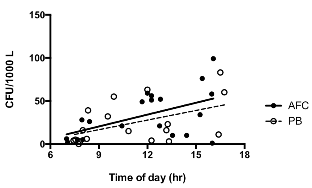
Total bacterial air counts rose during the day from <10 to 80–90 CFU/1000 L air both adjacent to the flow cytometer and at the nearby preparation bench (Figure 1). Airborne bacterial counts (CFU/1000 L air) in the clinical bacteriology laboratory spanned a wider range of values but not a significantly different distribution (Table 2). There was a significant fall in bacterial counts from air samples collected adjacent to the flow cytometer (54 CFU/1000 L air falling to 15.5 CFU/1000 L air) after a thorough laboratory clean. The comprehensive cleaning of laboratory surfaces and replacement of gowns was associated with a significant reduction in total air counts (Figure 2).
AFC, acoustic-enhanced flow cytometer; CML, clinical microbiology laboratory; ‘In use’ refers to AFC in operation for bacterial analysis; PB, preparation bench.
| Air samples | n | Lower limit | Median | Upper limit |
|---|---|---|---|---|
| AFC, not in use | 9 | 0 | 5 | 85 |
| In use AFC | 10 | 10 | 54 | 99 |
| Post clean AFC | 4 | 1 | 15.5 | 21 |
| PB | 20 | 0 | 15.5 | 83 |
| CML | 14 | 0 | 33.5 | 189 |
| All samples | 57 | 0 | 21 | 189 |
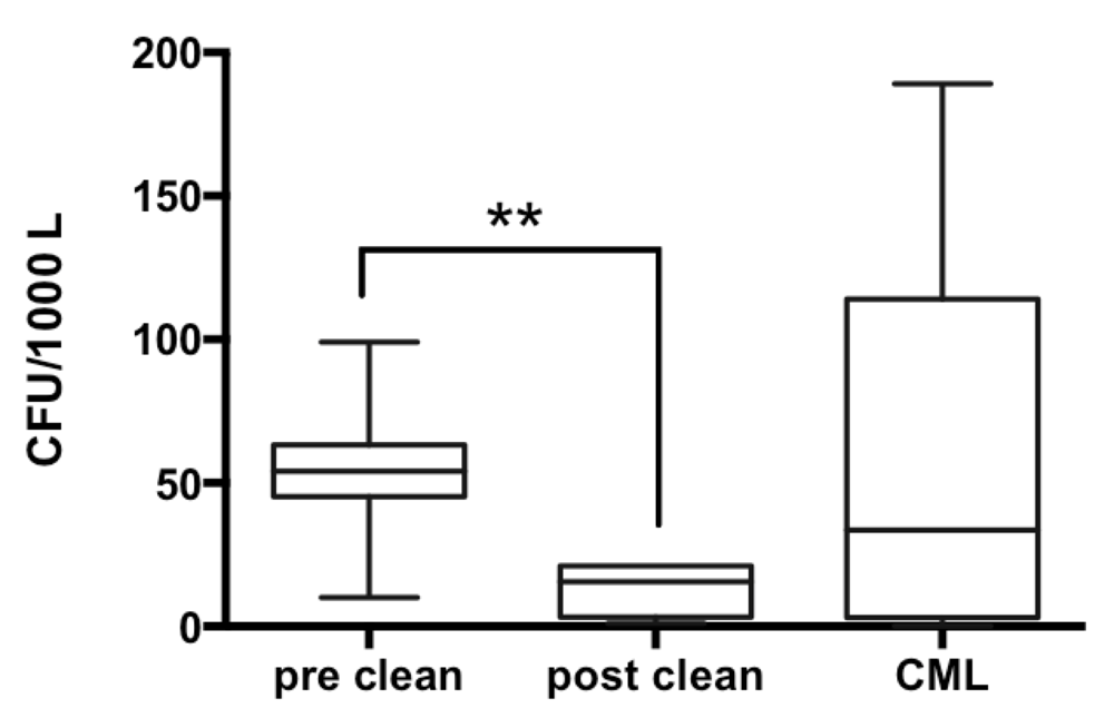
Pre clean research laboratory and post-clean median counts in close proximity to flow cytometer sample introduction port = 54 and 15.5 CFU/ 1000L, U = 2.5, p = 0.0090.
The stated aim of the present study was to assess the risk of personnel exposure to airborne bacteria potentially generated during analysis with an AFC, and determine any mitigation measures that would reduce any risk posed by airborne bacteria. The absence in air samples of any of the three bacterial species analysed with the flow cytometer over a month of intensive laboratory work indicates that these three species represent a low aerosolisation risk during the procedures we currently use. None of the analyses involve flow-assisted cell sorting, a process known to potentially generate bioaerosols12,14, since the flow cytometers in use in our laboratory do not have that capability. Until a similarly thorough analysis of bacterial fluorescence-activated cell sorting has been performed, the higher risk of bacterial aerosol generation by cell sorters should be considered when planning experiments and their corresponding biosafety measures. The absence of any of these three species on the external surfaces of the flow cytometer is further evidence to indicate that the cytometer sample introduction port does not easily generate bacterial aerosols and that basic procedures of wiping surfaces with 70% ethyl alcohol are sufficient to adequately maintain a safe working environment. While our data applies only to the three species we assessed and does not necessarily predict the likelihood of other bacteria becoming airborne during flow cytometer procedures, it is likely that the occupational risks of handling other bacterial species with the same physical properties can be assessed by this approach.
The dominance of skin commensal and environmental species, which has been reported before15,16, raises wider issues of industrial hygiene which have not been actively investigated in clinical bacteriology laboratories handling class 2 pathogens. Our air sampling data indicate that viable airborne bacteria are common and may reach high concentrations of colony forming units during peak laboratory working hours. These data do not distinguish between skin bacteria shed by laboratory staff and disturbance of those same species resting on inanimate surfaces, as these issues were beyond the scope of our investigation. Nevertheless, the reduction in airborne bacterial counts documented after a thorough cleaning of research laboratory surfaces and replacement of laboratory gowns suggests that dust particles may be a contributory factor. These sources may be a lesser concern in a clinical bacteriology laboratory, but in a research laboratory developing novel methods of analysing bacteria, airborne bacteria represent a potential source of media and equipment contamination that need to be brought under control. Contamination of the flow cytometer focussing, wash or other fluids even with non-biological particles is a source of background noise and needs to be avoided for optimal results. For this reason, bacterial flow cytometer analyses demand some of the contamination control discipline exercised in molecular diagnostic laboratories. The contamination control measures incorporated into FAST procedures include thorough 0.1 μm filtration of all fluids, flow cytometer analysis of suspending fluids for background particulate noise prior to bacterial analyses, sodium hypochlorite treatment of all flow cytometer effluent, and housing one of the acoustic flow cytometers inside a non-operating class 2 biosafety cabinet. Use of the non-operating cabinet gives the benefit of a physical barrier between the sample introduction port and the operator. Unfortunately, coarse vibration associated with safety cabinet operation may interfere with the accuracy of flow cytometer data capture. Housing the flow cytometer inside a static bio-bubble of suitable biocontainment level will cause less interference from vibration, when analysing biosafety containment level three (BL3) species.
This study showed that use of an AFC for bacterial analysis over extended periods did not contribute detectable concentrations of test bacteria to the population of bacteria we cultured from air samples collected in a research laboratory environment. Further, the majority of bacteria cultured were skin commensal and environmental bacteria, presumed to have been shed and dispersed or distributed in laboratory air by personnel movements during routine laboratory operation. The concentrations of airborne bacteria detected were comparable with those detected in a nearby clinical laboratory on the same biomedical campus and were significantly reduced after laboratory cleaning measures were introduced.
We undertook this work as part of a risk assessment of the hazards posed by analysing unfixed bacteria by flow cytometric methods. None of the bacteria being investigated by flow cytometry were detected in air samples, irrespective of whether the AFCs were in use. Furthermore, the levels of airborne bacteria detected in the research laboratory were lower than those detected in a large clinical bacteriology laboratory located in an adjacent building on the same campus. We conclude that our flow cytometric analyses of unfixed suspensions of K. pneumoniae, B. thailandensis and S. pneumoniae do not pose a risk to AFC operators or other personnel in the laboratory but caution against extrapolation of our results to other bacteria and/or different flow cytometric experimental procedures.
The data supporting the findings reported in this study have been uploaded to OSF: osf.io/z8uka17.
Dataset 1: FAST project bacterial air sampling. Quantitative air sampling data, times, locations, corresponding laboratory activities and qualitative bacteriologic identification results.
Datasets 2 – 6: FAST air sampling Table 1A, Table 1B, and Table 2, and Figure 1 and Figure 2. Analysis of bacterial air sampling during use of acoustic-enhanced flow cytometer for bacterial analysis and control settings.
Data are available under the terms of the Creative Commons Zero “No rights reserved” data waiver (CC0 1.0 Public domain dedication).
| Views | Downloads | |
|---|---|---|
| Gates Open Research | - | - |
|
PubMed Central
Data from PMC are received and updated monthly.
|
- | - |
Provide sufficient details of any financial or non-financial competing interests to enable users to assess whether your comments might lead a reasonable person to question your impartiality. Consider the following examples, but note that this is not an exhaustive list:
Sign up for content alerts and receive a weekly or monthly email with all newly published articles
Register with Gates Open Research
Already registered? Sign in
If you are a previous or current Gates grant holder, sign up for information about developments, publishing and publications from Gates Open Research.
We'll keep you updated on any major new updates to Gates Open Research
The email address should be the one you originally registered with F1000.
You registered with F1000 via Google, so we cannot reset your password.
To sign in, please click here.
If you still need help with your Google account password, please click here.
You registered with F1000 via Facebook, so we cannot reset your password.
To sign in, please click here.
If you still need help with your Facebook account password, please click here.
If your email address is registered with us, we will email you instructions to reset your password.
If you think you should have received this email but it has not arrived, please check your spam filters and/or contact for further assistance.
Comments on this article Comments (0)