Keywords
Systematic Review, pneumonia etiology, bacterial pneumonia, children
Systematic Review, pneumonia etiology, bacterial pneumonia, children
Globally, pneumococcal infections, caused by Streptococcus pneumoniae (Pneumococcus), are one of the leading causes of morbidity and mortality in children <5 years of age1. A variety of clinical syndromes of varying severity are associated with pneumococcus, including pneumonia, meningitis, bacteremia, otitis media and sinusitis2. It has been estimated that prior to the introduction of pneumococcal conjugate vaccines (PCVs), diseases caused by pneumococcus were responsible for approximately 600,000 deaths per year globally in children 1-59 months of age3.
Pneumonia is among the leading causes of mortality in children under 5 years of age1,4. The main causative pathogens attributable to pneumonia include Streptococcus pneumoniae, Haemophilus influenzae type B, all of which have vaccine-preventable bacterial causes, and respiratory syncytial virus5. Infants and young children are at highest risk for serious disease6, with children younger than 4 months being more likely to die7. In addition to pneumococcus, a variety of other infectious agents are related to pneumonia in children.
In the last two decades, more than 140 countries globally have introduced PCVs into national routine immunization schedules. Several studies have demonstrated the impact of PCVs on reducing invasive pneumococcal diseases and hospitalizations due to pneumonias2,8,9. However, pneumonia mortality is the greatest concern for policymakers and donors, and there is limited evidence on the impact of PCVs on pneumonia deaths in children.
Recent evidence from countries in Latin American using secondary mortality data demonstrated the impact of PCVs on pneumonia mortality in children under 5 years of age10–13. Most studies did not include children <3 months of age assuming that perinatal causes of mortality and other etiologic agents and not pneumococcal disease are responsible for the pneumonia mortality in this age group. Nonetheless, this assumption is not fully backed up by the very little available evidence in the literature on the etiology of pneumonia in this age group. Although selected studies have indicated that respiratory viruses are the most common pathogens of pneumonia in infants and toddlers, some investigators have implicated pneumococcus and Haemophilus in 4–20% of cases. These findings vary significantly in developing versus industrialized countries, over time, and depending on laboratory methods used to assess etiologies.
It is still not clear whether pneumococcus is a significant cause of pneumonia in younger children, particularly neonates and children <3 months of age. Whether or not to include children in this age group in impact assessment studies will depend on evidence suggesting whether pneumococcus is a significant etiology of pneumonia and thus an important burden in children under 3 months of age.
This systematic review aims at summarizing the evidence of the bacterial etiology of respiratory infections in children under 3 months of age, in particular the role of pneumococcus as a significant etiology in this age group.
The study protocol was published in PROSPERO under registration number CRD42020158091. We followed PRISMA recommendations14, and a completed PRISMA checklist is provided as extended data15.
A systematic literature review was performed to identify all available data from published studies on the etiology of bacterial pneumonia in children younger than 3 months of age. Electronic searches were conducted in the following databases: Medline/PubMed, Embase, Central and Index Medicus Global (including Lilacs-Latin America and Caribbean, Regional Index Medicus (IM), including IM Western Pacific (WPRIM), IM Africa (AIM), IM South-East Asia (IMSEAR) and IM East Mediterranean (IMEMR). A complementary search was conducted in the electronic library SciELO and in Scholar. Additionally, references of selected articles and reviews were screened. No date, location, or language limits were placed on the searches of publications through April 30th, 2021. Detailed search strategies for each database are presented in the extended data15.
Studies reporting primary data about respiratory infections or invasive bacterial disease/sepsis secondary to pneumonia in children under 3 months of age of both sexes, regardless of any co-morbidity, were considered. We included studies that reported on the following syndromes as disease outcomes: bacterial pneumonia, pneumonia (clinical or X-ray confirmed) pneumonitis/bronchitis, Acute respiratory illness (ARI), pulmonary complications, deaths due to pneumonia, respiratory infections, severe or hospitalized pneumonia, community acquired pneumonia (CAP), para-pneumonic pleural effusion (PPE) and/or bloodstream infection/sepsis secondary to pneumonia.
Any etiologies assessed by laboratory, considering any diagnostic method and a variety of clinical specimens were incorporated.
We included citations reporting on primary studies in which the etiology of bacterial pneumonia or invasive bacterial disease secondary to bacterial pneumonia is assessed, including mostly observational studies (descriptive studies, case series, case-control, cohort and cross-sectional studies), but also randomized controlled trials (RCTs). Case series were included only if at least 10 cases are reported in the target age-group.
We excluded studies which did not report or did not provide data for the specific age subgroup of our interest, studies which did not report on laboratory confirmed etiology for the outcome of interest, and studies that reported on infections secondary to other non-respiratory primary focus (or which primary focus was unknown).
Case reports, guidelines/recommendations, letters and reviews were excluded. Also, laboratory studies in which the clinical syndrome is not described, and case series with less than 10 events are reported were excluded.
Studies evaluating etiology of the following syndromes/diagnosis were excluded: hospital-acquired pneumonia, necrotizing pneumonia, aspirative pneumonia, pneumocystosis, interstitial pneumonia, influenza like illness, and bronchiolitis. Also, studies evaluating an outbreak of group of cases with a specific etiologic agent already defined (ii.e. adenovirus outbreak) were also excluded. Finally, studies evaluating laboratory samples (not children with clinical syndromes) and carriage studies were excluded.
Citations retrieved in bibliographic searches were uploaded in EndNote 20 reference manager, and deduplication were performed. Remaining citations were screened by four independent reviewers (CT, MQ, MTV, MSM) in the first step, where titles and abstracts were reviewed for inclusion criteria. Screened articles were categorized as potentially eligible, unclear, or excluded. Citations on which the pair of reviewers disagreed were discussed or assessed by a third reviewer. Full text of papers meeting inclusion criteria and those unclear were obtained. In the second step, full texts were read and assessed for information on whether they meet inclusion criteria by four reviewers (CT, MQ, MTV, MTCO) and disagreements were resolved by discussion. In this step articles were categorized as included, excluded, or uncertain. Studies were categorized as uncertain when through full review we were not able to extract the information on etiologic agents of pneumonia for the specific ≤2 month of interest, because reported results were aggregated in larger age groups. For these cases, we contacted the authors of all original studies which were published in or after 2015. The rationale was that for more recent studies, the authors might have information on etiologies for the specific age subgroup of interest, even though these were not depicted in the publication. After receiving author’s response, if data were obtained, articles were included but if authors didn’t answer or didn’t have the data, articles were excluded. One additional round of deep full text analysis was done with the complete list of selected studies resulting in new exclusions according to inclusion criteria.
Data extraction was done by five independent reviewers (CT, MQ, MTV, MSM, MTCO), using abstraction forms developed specifically for this systematic review.
To avoid multiple counting of reports from the same study, citations from the same study group on data originated from the same study protocol, population or information system were grouped for extraction, and reported as a single study.
Data extracted included: country; year of publication; study design; study period; sample size; demographic information (average age, sex, ethnicity); diagnostic criteria; laboratory method for diagnosis and etiologic agent; laboratory specimen considered for diagnosis; outcome definition; secondary outcomes; availability of data for ≤2 months of age; number of study subjects; number and proportion of etiologic agents by each group of etiologies.
Quality assessment of studies included in the review was conducted using the JBI critical appraisal checklist for cross-sectional, case-control, cohort, prevalence, and case series studies.
A descriptive analysis of study characteristics including study design, respiratory syndromes/outcomes considered, biological specimen evaluated, and laboratory method used for etiologic confirmation was conducted. For all studies, the main measure of interest was the etiologic agents identified. Descriptive data on the etiologies of respiratory infections in children under 3 months of age was analyzed and are presented as percentages. As a variety of syndromes, biological specimens, and laboratory diagnostic methods were reported in the various studies, we present the results stratified by diagnostic method.
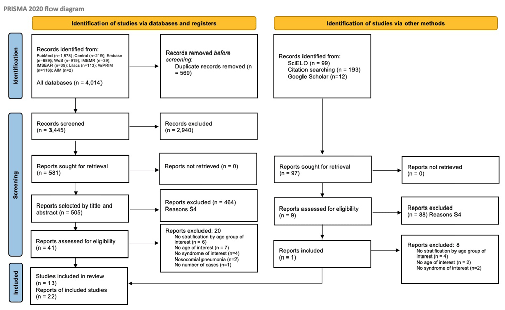
A total of 4,313 studies were retrieved in searches. After eliminating duplicates, a total of 3,744 references were screened by title and abstract review. Out of the 602 selected citations, a further 580 were excluded in two rounds of review, with 22 remaining papers eligible for data extraction reporting on 13 studies included in this review (Figure 1). A complete list of reasons for excluding studies as well as references are provided in the extended data. Out of correspondences to authors of 54 studies, 16 responses were received, but new data was obtained for only 9 studies, which are included in 22 selected papers. Detailed list of included papers by database can be found in the extended data15.
13 (n=13) studies were considered in this review (Table 1). Studies range over four decades (1980-2020), present different study designs, and consider a wide variation of number of children enrolled and assessed. Despite target age group being younger than 3 months of age, two studies evaluated only neonates aged ≤28 days, while some studies evaluated children <3 months including neonates and others excluded neonates from the study, thus including only children >28 days to <3 months.
| Characteristics | n | % |
|---|---|---|
| Study period | ||
| 1980-2000 | 5 | 38.5% |
| 2000-2010 | 3 | 23.0% |
| 2010-2020 | 5 | 38.5% |
| Study design | ||
| Prospective cohort | 7 | 53.8% |
| Case control | 1 | 7.7% |
| Retrospective cohort | 3 | 23.1% |
| Case series | 1 | 7.7% |
| Cross-sectional | 1 | 7.7% |
| Sample size | ||
| 10 – 49 children | 8 | 61.5% |
| 50 – 150 children | 1 | 7.7% |
| 150 children and over | 4 | 30.8% |
| Age groups | ||
| Only neonates (≤28 days) | 2 | 15.4% |
| <3 months (including neonates) | 5 | 38.5% |
| >28 days to <3 months | 6 | 46.2% |
| Clinical syndromes considered | ||
| Hospitalized community acquired pneumonia | 7 | 53.8% |
| Acute Respiratory illness (ARI) | 1 | 7.7% |
| Sepsis secondary to pneumonia | 5 | 38.5% |
| Biological specimen* | ||
| Blood | 12 | 92.3% |
| Nasal/throat swabs | 8 | 61.5% |
| Broncho-alveolar lavage (BAL) | 1 | 7.7% |
| Pleural effusion/aspirate | 3 | 23.1% |
| Lung biopsy | 1 | 7.7% |
| Urine | 3 | 23.1% |
| Cerebrospinal fluid (CSF) | 5 | 38.5% |
| Etiologic groups evaluated | ||
| Bacteria only | 3 | 23.1% |
| Virus only | 1 | 7.7% |
| Virus and Bacteria | 9 | 69.2% |
| Diagnostic methods used* | ||
| Culture | 12 | 92.3% |
| PCR/molecular | 6 | 46.2% |
| Serology | 5 | 38.5% |
| Antigen tests | 2 | 15.4% |
While one study only assessed viral etiologies16, three studies evaluated only bacterial etiologies17–20. Most studies report on blood (n=12) and nasopharyngeal swabs/aspirates or nasal washing (n=8), although some studies also collected other specimens, including cerebrospinal fluid (CSF), urine, broncho-alveolar lavage (BAL), pleural effusion and lung biopsies. Added to this variability of samples available and tests conducted, there were varying methods used and for etiologic diagnosis, with most studies reporting cultures, but some also using serology and antigen testing for viral infections. Most recent studies16,21–33 included molecular techniques, with known increased sensitivity (i.e., ability to detect pathogens) to identify many etiologic agents. The variety of study designs and methods used in the studies made it inappropriate to conduct a meta-analysis.
Etiologies identified by each study have, as expected, varied greatly considering study characteristics and methods as described above. In Table 2 below, main study characteristics and etiologic agents identified are presented (Table 2).
| Author, year of publication | Location, study period | Type of syndrome | Age group | Specimen | Lab methods | Number of children evaluated by laboratory | Bacterial etiology (%) | Etiology results (number of chilren with laboratory confirmed etiology, by etiologic agent) |
|---|---|---|---|---|---|---|---|---|
| Misra, S (1991)34 | India (1986-87) | Hospitalized community acquired pneumonia with PPE | Neonates (<28 days) | Blood and Lung Aspirates | Culture, antigen test and serology for virus and bacteria | 44 | 22 (5.7%) | 10 S. pneumoniae 15 Gram-negatives 02 Streptococcus 04 S. epidermidis 01 Coagulase-negative Staphylococcus |
| The WHO Young Infant Study Group (1999)35 | Ethiopia, Papua New Guinea, Gambia, Philippines (1991-93) | Severe X-ray confirmed community acquired pneumonia and/or sepsis | <3 months | Blood, urine, NPA and CSF | Culture, viral immunofluorescence for virus and bacteria | 2,452 children with blood cultures | 167 positive blood cultures | 33 S. pneumoniae 34 S. aureus 29 S. pyogenes 02 Streptococcus group B 02 Streptococcus groups D/E/F 02 Streptococcus group G 17 H. influenzae 41 Gram-negatives |
| Muhe, L (1999)36 | Ethiopia (1991-93) | Severe X-ray confirmed community acquired pneumonia and/or sepsis | <3 months | Blood, NPA and CSF | Culture, viral immunofluorescence for virus and bacteria | 405 of which 202 NPA for viral etiologies and C. trachomatis | 13 pneumonia and 31 sepsis by culture | Pneumonia (n=13) 05 S. pneumoniae 03 H. influenzae 09 S. pyogenes 05 Salmonella spp 10 E. coli 02 S. aureus 02 Other Gram-negatives Sepsis (n=31) 08 S. pneumoniae 02 H. influenzae 08 S. pyogenes 02 Salmonella spp 08 E. coli 02 S. aureus 01 Gram-negative Viral Etiologies 57/202 (28%) RSV 32/202 (16%) C. trachomatis |
| Lehman D (1999)37,38 | Papua New Guinea (1991-93) | Severe X-ray confirmed community acquired pneumonia and/or sepsis | <3 months | Blood, NPA and CSF | Culture, viral immunofluorescence for virus and bacteria | 845 | 48 | 13 S. pneumoniae 13 S. pyogenes 10 S. aureus 03 E.coli 03 Enterococcus faecalis 02 H. influenzae 02 K. pneumoniae 01 S. agalactiae 01 Streptococcus group G 01 Enterobacter cloacae |
| The PERCH Study21,24,25,31–33 | PERCH – Kenya, Gambia, Mali, Zambia, South Africa, Thailand and Bangladesh (2011-14) | Severe X-ray confirmed community acquired pneumonia | >28 days to <3 months | Blood, NPA, urine, BAL, PE, lung aspirates, gastric aspirates | Culture, PCR, serology, Antigen tests for virus and bacteria | 810 with blood cultures | 349 culture positive, of which 10 bacterial | 02 S. pneumoniae 02 H. influenzae 03 S. aureus 01 Salmonella spp 02 other Enterococcus and Streptococcus |
| Rhie, K (2018)19 | Korea (2006-10) | Invasive bacterial infection secondary to pneumonia in hospitalized children | < 3months | Blood, PPE, CSF | Culture for bacteria only | 113 with positive cultures | Only tested for bacteria | 27 S. aureus 18 E. coli 55 S. agalactiae |
| Lee, JH (2011)18 | Korea (1996-2005) | Invasive bacterial infection secondary to pneumonia in hospitalized children | < 3months | Blood, PPE, CSF | Culture for bacteria only | 95 (in all age groups) | 13 (13.7%) | 07 S. aureus 02 S. pneumoniae 04 S. agalactiae |
| Wang, H (2010)20 | China (2006-08) | Hospitalized community acquired pneumonia | Neonates (<28 days) | Blood (sample of positive sputum samples) | Culture for bacteria only | 80 with positive blood cultures | Only tested for bacteria | 38 K. pneumoniae 20 E. coli 16 S. aureus 06 S. epidermidis |
| Finianos, M (2019)16 | Lebanon (2013-14) | Hospitalized respiratory infections | >28 days to <3 months | NPA | PCR for viruses only | 25 | Only tested for virus | 12 RSV 05 Rhinovirus 03 Bocavirus 02 Influenza 01 Coronavirus |
| Nascimento- Carvalho, CM (2011, 2013, 2015, 2019)26–29 | Brazil (2003-05) | Hospitalized community acquired pneumonia | >28 days to <3 months | Blood and throat NPA | Culture, PCR, serology for virus and bacteria | 16 | 12 (75%) | 06 C. trachomatis 01 Rhinovirus 01 Parainfluenza 03 RSV + C. trachomatis 01 RSV + S. pneumoniae + C. trachomatis 01 Parainfluenza + C. trachomatis 01 Enterovirus + S. pneumoniae + C. trachomatis 01 Rhinovirus + human metapneumovirus |
| Jullien, S (2020)23 | Bhutan (2017-18) | Hospitalized X-ray confirmed community acquired pneumonia | >28 days to <3 months | Blood and NP washing | Culture, PCR, Antigen tests for virus and bacteria | 13 (12 with culture and 9 with NP washing) | 1 (8%) | 03 RSV 02 Rhinovirus 01 RSV + Rhinovirus 01 Parainfluenza + Rhinovirus |
| Nathan, AM (2020)30 | Malaysia (2014-16) | Severe X-ray confirmed community acquired pneumonia | >28 days to <3 months | Blood and induced sputum | Culture, PCR, and immunofluorescence for virus and bacteria | 45 | 24 (48%) | 08 S. aureus 06 H. influenzae 02 S. pneumoniae 02 S. pneumoniae + rhinovirus 02 S. aureus + rhinovirus 02 S. aureus + RSV 01 H. influenzae + rhinovirus 01 H. influenzae + RSV 02 Rhinovirus 02 Metapneumovirus 01 Bocavirus 01 RSV 01 Influenza A |
| Gareca Perales, J (2021)22 | Bolivia (2016-17) | Hospitalized community acquired pneumonia | >28 days to <3 months | Blood and nasal washing | Culture and PCR for virus and bacteria | 47 | 5 (11%) | 12 RSV 05 RSV + Rhinovirus 04 Rhinovirus 02 Influenza 02 B. pertussis 02 S. aureus 01 S. pneumoniae 01 CMV 01 Enterovirus |
The only study conducted in the 1980s34 in India, evaluated only neonates up to 28 days. This study of 44 neonates reports no Streptococcus agalactiae as an aetiologic agent of neonatal pneumonia, but rather demonstrates a high proportion of S. pneumoniae (22.4%) (using antigen testing), and Gram-negative agents (25%).
A multinational World Health Organization (WHO) study in three countries to assess etiology of severe disease in children < 3 months of age, considering also but not restricted to pneumonia, was conducted from 1991 to 199335. This study is reported in different papers, one of which describes results in all countries combined35, and country specific results, namely Ethiopia36 and Papua New Guinea37,38, as there are some variations in methods in each study site. This was the largest prospective study of early infant infections in developing countries, and it also reported on the absence of Streptococcus agalactiae and the importance of Gram-positive (61%) and Gram-negative (24%) organisms among the 167 positive blood cultures with isolates identified. Among these, S. pneumoniae, S. aureus and S. pyogenes (Gram-positives), and E. coli and Salmonella (Gram-negatives) are the most noteworthy. Viral etiologies were also important pneumonia agents, as reported in Ethiopia36.
A retrospective laboratory-based surveillance study including children with bacterial invasive disease was conducted in Korea, and two papers report on different study periods, from 1996-200518, and from 2006-201019. This study only considered bacterial etiologies from blood, PPE, CSF, and here S. agalactiae is frequently isolated in both study periods.
Another study evaluating only neonates up to 28 days of age in China from 2006-200820 also demonstrated a significant proportion of Gram-negative agents in children hospitalized with community acquired pneumonia, mostly K. pneumonia and E. Coli.
One study evaluated viral etiologies of hospitalized children with respiratory infections only16. Children aged up to 28 days were excluded, so only children aged over 28 days and under 3 months of age were considered. Molecular methods were used to assess etiologies in nasopharyngeal aspirates (NPA) (no other specimen was obtained and evaluated). A high proportion of respiratory syncytial virus (RSV) was observed in children with positive isolates.
Nascimento-Carvalho et al.26–29 in multiple prospective cross-sectional studies conducted in Brazil during 2003–2005 and evaluating 19 different etiologies in blood and NPA using culture, serology and PCR, report the importance of viral etiologies in children hospitalized with community acquired pneumonia. Here, children aged 28 days and younger were excluded from the study, so data presented is for children from 1 to 3 months of age.
Finally, a large WHO multinational study in seven countries to assess the etiology of severe pneumonia, using a case-control design and including hundreds of children, was conducted from 2011-201421,24,25,31–33. Children aged 28 days and younger were also excluded, and various specimen types and multiple laboratory methods were used to identify viral and bacterial etiologies of severe pneumonia. Findings also reinforce the importance of viral etiologies in children aged 1–3 months, but bacterial agents, particularly S. pneumoniae and S. aureus, were also reported as relevant agents.
More recent studies have similar methods, including prospective cohort designs, the use of molecular methods, and collection of various specimens including blood and NPA at a minimum22,23,30. While the studies in Bhutan23 and Bolivia22 reported a significant proportion of viral etiologies, particularly RSV and rhinovirus, the study in Malaysia30 reported a high proportion of Gram-positive bacterial agents, particularly S. aureus, H. influenzae, and S. pneumoniae, isolated or combined with viral etiologies.
The risk of bias assessment of the studies is presented in Table 3. In general, studies presented high risk of bias, particularly due to design, small number of subjects, specimens collected, and laboratory methods. Many of them were conducted in different decades, when availability and accuracy of diagnostic tools varied significantly. Very few studies included controls, namely the multinational WHO Young Infant Study Group (1999)35, and PERCH Study21,24,25,31–33. Given the paucity of evidence, we opted to report on all studies, and consider their limitations and potential biases in interpreting the results.
| Author, year of publication | Location, study period | Study design | Quality assessment |
|---|---|---|---|
| Misra, S (1991)34 | India (1986-87) | Case series | Fair |
| The WHO Young Infant Study Group (1999)35 | Ethiopia, Papua New Guinea, Gambia, Philippines (1991-93) | Case control | Excellent |
| Muhe, L (1999)36 | Ethiopia (1991-93) | Case control | Excellent |
| Lehman D (1999)37,38 | Papua New Guinea (1991-93) | Case control | Excellent |
| The PERCH Study21,24,25,31–33 | PERCH – Kenya, Gambia, Mali, Zambia, South Africa, Thailand and Bangladesh (2011-14) | Case control | Excellent |
| Rhie, K (2018)19 | Korea (2006-10) | Retrospective cohort | Fair |
| Lee, JH (2011)18 | Korea (1996-2005) | Retrospective cohort | Fair |
| Wang, H (2010)20 | China (2006-08) | Retrospective cohort | Fair |
| Finianos, M (2019)16 | Lebanon (2013-14) | Prospective cohort | Fair |
| Nascimento-Carvalho, CM (2011, 2013, 2015, 2019)26–29 | Brazil (2003-05) | Cross sectional | Good |
| Jullien, S (2020)23 | Bhutan (2017-18) | Prospective cohort | Good |
| Nathan, AM (2020)30 | Malaysia (2014-16) | Prospective cohort | Good |
| Gareca Perales, J (2021)22 | Bolivia (2016-17) | Prospective cohort | Good |
Pneumonia is a very frequent childhood disease leading to disease burden significantly higher in children when compared to other age groups. Several studies, many of which conducted in developing countries, have evaluated pneumonia etiologies in the past decades. Understanding pneumonia etiology is key to guiding diagnosis and management approaches to pediatric pneumonia.
It is well known that pneumonia etiology varies by age. Nonetheless, very limited evidence is available for young children, particularly children under 3 months of age. Determining the etiology of community acquired pneumonia in children, including severe disease leading to hospitalizations, is very important not only to define treatment guidelines, but also to implement preventive strategies at national level, and more recently to also assess the impact of selected interventions, as pneumococcal conjugate vaccines (PCVs) are introduced.
Identifying the cause of pneumonia in children is difficult because of varying syndromic presentations, challenges in obtaining specimens for laboratory assessment, and the lack of rapid, commercially available, accurate laboratory tests for most pathogens, among others. A recent landscape assessment and literature review conducted by Gilani et al.39 reported that published or ongoing (at the time) studies of pneumonia etiology in children present a multiplicity of case definitions, levels of clinician involvement, facility types, specimen collection, and laboratory techniques, thus reinforcing the need for the standardization of methods and analyses of pneumonia etiology in children.
Limited reviews have reported on the etiology of pneumonia in children, mostly in specific locations and in younger than 5 years of age40. Studies conducted in developed countries clearly demonstrate that the pattern of etiologic agents causing pneumonia in children, in particular severe pneumonia, has been changing over the past decades. While in the 1980s bacterial agents including Staphylococcus bacteria (aureus and pyogenes) were the main causative agents of severe pneumonia in children, over time studies began reporting an increase in the proportion of Gram-negative agents and Group B Streptococcus (S. agalactiae), which accounted for most pneumonia cases in children. Also, there is growing evidence demonstrating the importance of viral etiologies, including RSV, rhinovirus, influenza, parainfluenza, alone or in combination with bacterial pathogens, as important etiologies of pneumonia in children.
In the Canadian Guidelines for treatment of pediatric pneumonia from 199717, the reported main pathogens causing pneumonia in infants aged 1-3 months are, in order of frequency: Chlamydia trachomatis, RSV, other respiratory viruses, and Bordetella pertussis.
In a review article published in 2002, McIntosh5 reports on the bacterial and viral agents causing pneumonia in children, particularly S. pneumoniae and S. pyogenes, S. aureus and H. influenza among bacterial agents, and RSV, influenza, parainfluenza, adenovirus, and rhinovirus among viral agents. McIntosh5 reinforces that for treatment decision making, one should first consider the age of the child. To that end, no comprehensive review has been conducted on the etiology of pneumonia in children under 3 months of age.
This systematic review included 13 studies reported in 22 publications, conducted from 1986 through 2020 in a variety of locations, mainly in developing countries. Results were variable, depending on time in which study was conducted, study design, and laboratory methods used.
Earlier studies conducted in the 80s and 90s34–37 demonstrate that S. pneumoniae is a very important etiologic agent even in neonates. Viral etiologies including RSV, influenza and parainfluenza were also observed in the WHO multicenter prospective study35–37.
A retrospective study in Korea18,19 conducted over 1996 to 2010 reported S. agalactiae as a significant agent. Nonetheless, these studies were severely biased for various reasons. First, the study was retrospective and based on laboratory surveillance, with no clinical information of patients enrolled, but rather considering invasive disease as of pulmonary focus when pulmonary or pleural specimens had been obtained. In addition, this study included both community and nosocomial infections, it not being possible to disaggregate them. Finally, only bacteria were evaluated and no viral etiologies. This study also included children younger than 28 days.
Another retrospective cohort study conducted in China20 evaluated only bacterial etiology on young neonates aged ≤28 days. Furthermore, only sputum specimens were collected and processed, which imposes major biases in this study as well.
More recent research conducted in the past 15 years, using prospective cohort designs and better standardized methods and case definitions, and applying molecular diagnostic techniques to evaluate etiologic agents including bacterial and viral etiologies, suggests a significant proportion of viral agents causing pneumonia in children younger than 3 months of age16,21–32,33. Of note, Finianos et al.16 in Lebanon evaluated viral agents only. Nathan et al.30 in Malaysia also report a significant proportion of bacterial etiologies, particularly S. pneumonia, H. influenza and S. aureus. This was also reported by the most robust body of evidence to date on the etiology of hospitalized pneumonia in children, resulting from a WHO multinational cohort study conducted in 7 study sites from 2011-2014, the Pneumonia Etiology Research for Child Health (PERCH) study21,24,25,31–33.
This review demonstrates that available evidence on etiology of pneumonia in young children, particularly children younger than 3 months of age is based on a variety of studies with non-standardized methodology. Syndromes and case definitions as well as age subgroups included (younger than 7 days and younger than 28 days) vary significantly among studies. Samples collected and tests performed also vary significantly, and also over time, with molecular methods available in more recent studies. Studies also vary in terms of sample size, and time and locality in which it has been conducted. All of these are known factors which may influence the reported etiology and also the ability to identify selected agents. Adequate specimens and testing methods should be used for studies evaluating etiology of pneumonia in children, in particular molecular techniques with higher sensitivity.
Despite the above limitations and challenges, this review reinforces that Gram-positive organisms, in particular S. pneumoniae, are still important etiologic agents of pneumonia in children under 3 months of age and should thus be considered when assessing impact of PCV in the children. In addition, viral etiologies are also important, responding for a significant proportion of pneumonia in children younger than 3 months of age.
All data underlying the results are available as part of the article and no additional source data are required.
Harvard Dataverse: Bacterial etiology of pneumonia in children up to 2 months of age: a systematic review. https://doi.org/10.7910/DVN/GIYVPD15
This project contains the following files:
Harvard Dataverse: PRISMA flowchart and checklist for "Bacterial etiology of pneumonia in children up to 2 months of age: a systematic review". https://doi.org/10.7910/DVN/GIYVPD
Data are available under the terms of the Creative Commons Zero "No rights reserved" data waiver (CC0 1.0 Public domain dedication).
| Views | Downloads | |
|---|---|---|
| Gates Open Research | - | - |
|
PubMed Central
Data from PMC are received and updated monthly.
|
- | - |
Provide sufficient details of any financial or non-financial competing interests to enable users to assess whether your comments might lead a reasonable person to question your impartiality. Consider the following examples, but note that this is not an exhaustive list:
Sign up for content alerts and receive a weekly or monthly email with all newly published articles
Register with Gates Open Research
Already registered? Sign in
If you are a previous or current Gates grant holder, sign up for information about developments, publishing and publications from Gates Open Research.
We'll keep you updated on any major new updates to Gates Open Research
The email address should be the one you originally registered with F1000.
You registered with F1000 via Google, so we cannot reset your password.
To sign in, please click here.
If you still need help with your Google account password, please click here.
You registered with F1000 via Facebook, so we cannot reset your password.
To sign in, please click here.
If you still need help with your Facebook account password, please click here.
If your email address is registered with us, we will email you instructions to reset your password.
If you think you should have received this email but it has not arrived, please check your spam filters and/or contact for further assistance.
Comments on this article Comments (0)